Anti-AIF-1/Iba1 Rabbit mAb - Microglia Marker
Catalog number :AT0682
Apoptosis-inducing factor (AIF, PDCD8) is a ubiquitously expressed flavoprotein that plays a critical role in caspase-independent apoptosis. AIF is normally localized to the mitochondrial intermembrane space and released in response to apoptotic stimuli. Treatment of isolated nuclei with recombinant AIF leads to early apoptotic events, such as chromatin condensation and large-scale DNA fragmentation. Studies of AIF knockout mice have shown that the apoptotic activity of AIF is cell type and stimuli-dependent. Also noted was that AIF was required for embryoid body cavitation, representing the first wave of programmed cell death during embryonic morphogenesis. Structural analysis of AIF revealed two important regions, the first having oxidoreductase activity and the second being a potential DNA binding domain. While AIF is redox-active and can behave as an NADH oxidase, this activity is not required for inducing apoptosis. Instead, recent studies suggest that AIF has dual functions, a pro-apoptotic activity in the nucleus via its DNA binding and an anti-apoptotic activity via the scavenging of free radicals through its oxidoreductase activity.
- Overview
- Reactivity
- Human, Mouse, Rat, Monkey
- Tested applications
- Western Blotting 1:1000Immunoprecipitation 1:200Immunohistochemistry (Paraffin) 1:500Optimal dilutions/concentrations should be determined by the end user.
- Specificity
- This antibody detects endogenous levels of total AIF/1Iba1 protein.
- Properties
- Immunogen
- Recombinant fragment, corresponding to a region within acids 1-147 of Human Iba1 (UniProt: P55008).
- Clonality
- Monoclonal, clone number: 4G2
- Isotype
- Rabbit IgG
- Form
- Liquid, 100 μl,1mg/ml, PBS (pH 7.2) and 40% Glycerol,0.02% Sodium Azide
- Storage instruction
- Store at +4°C short term (1-2 weeks). Aliquot and store at -20°C, Avoid freeze / thaw cycle.
- Host
- Rabbit
- Applications
- WB Image

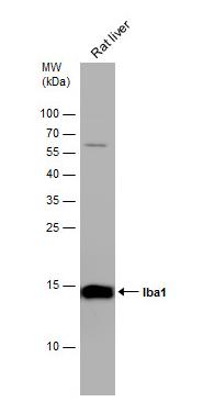
Western Blot: AIF-1/Iba1 Antibody - Rat tissue extract (50 ug) was separated by 15% SDS-PAGE, and the membrane was blotted with Iba1 antibody diluted at 1:1000. The HRP-conjugated anti-rabbit IgG antibody was used to detect the primary antibody.
- IHC Image

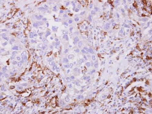
Immunohistochemistry-Paraffin: AIF-1/Iba1 Antibody - Paraffin-embedded breast cancer stroma. Iba1 antibody dilution: 1:500.
- ICC/IF Image

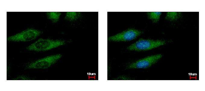
Immunocytochemistry/Immunofluorescence: AIF-1/Iba1 Antibody - HeLa cells were fixed in iced-cold MeOH for 5 min. Green: AIF1 protein stained by Iba1 antibody diluted at 1:500. Blue: Hoechst 33343 staining.
Related Products
Reviews
loading...