Anti-APC Rabbit mAb
Catalog number :AT0391
Tumor suppressor. Promotes rapid degradation of CTNNB1 and participates in Wnt signaling as a negative regulator. APC activity is correlated with its phosphorylation state. Activates the GEF activity of SPATA13 and ARHGEF4. Plays a role in hepatocyte growth factor (HGF)-induced cell migration. Required for MMP9 up-regulation via the JNK signaling pathway in colorectal tumor cells. Acts as a mediator of ERBB2-dependent stabilization of microtubules at the cell cortex. It is required for the localization of MACF1 to the cell membrane and this localization of MACF1 is critical for its function in microtubule stabilization.
- Overview
- Description
- Rabbit monoclonal antibody to APC
- Reactivity
- Mouse, Human, Xenopus laevis
- Tested applications
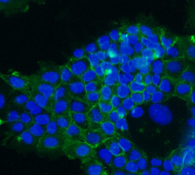
- ICC/IF: Use a concentration of 1 µg/ml.
WB: Use at an assay dependent dilution. Predicted molecular weight: 312 kDa.
IP: Use at an assay dependent dilution.
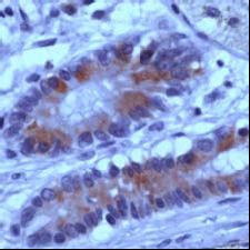
IHC-P: 1/100. Perform heat mediated antigen retrieval before commencing with IHC staining protocol.
- Properties
- Immunogen
Synthetic peptide (the amino acid sequence is considered to be commercially sensitive) (Human)(C terminal)
- Clonality
- Monoclonal, clone number: 2G7
- Isotype
- IgG
- Form
- Liquid
Preservative: Sodium Azide
Constituents: BSA, 10mM PBS, pH 7.4
- Storage instruction
- Shipped at 4°C. Upon delivery aliquot and store at -20°C. Avoid freeze / thaw cycles.
- Database links
- Entrez Gene: 324 Human
- Entrez Gene: 11789 Mouse
- Entrez Gene: 24205 Rat
- Entrez Gene: 399455 Xenopus laevis
- Omim: 611731 Human
- SwissProt: P25054 Human
- SwissProt: Q61315 Mouse
- SwissProt: P70478 Rat
- Applications
- IHC Image

Normal human colon section stained with AT0391 (1:100 for 10 min at RT). Sections were pre-treated by boiling in 10mM citrate buffer, pH 6.0 for 10 min followed by cooling at RT for 20 min.
- ICC/IF Image

ICC/IF image of AT0391 stained A431 cells. The cells were 4% paraformalehyde fixed (10 min) and then incubated in 1%BSA / 10% normal goat serum / 0.3M glycine in 0.1% PBS-Tween for 1h to permeabilise the cells and block non-specific protein-protein interactions. The cells were then incubated with the antibody (AT0391, 1µg/ml) overnight at +4°C. The secondary antibody (green) was DyLight® 488 goat anti-rabbit IgG (H+L) used at a 1/250 dilution for 1h. Alexa Fluor® 594 WGA was used to label plasma membranes (red) at a 1/200 dilution for 1h. DAPI was used to stain the cell nuclei (blue) at a concentration of 1.43µM.
Related Products
Reviews
loading...