CoIP, ChIP, CUT&RUN, CUT&Tag Expert! My Basket (...)
Worldwide delivery!
Having the Best Alpaca Nanobody Products!
Home | About us | Contact us | How to order |
    My Base Info
    Shopping Basket
    My Orders
    My Shipping Info
    Modify Password
    Log Out

    Anti-ATPB Rabbit mAb - Mitochondrial Marker

    Catalog number :AT0811
    loading...
    Background: 
    Mitochondrial membrane ATP synthase (F(1)F(0) ATP synthase or Complex V) produces ATP from ADP in the presence of a proton gradient across the membrane which is generated by electron transport complexes of the respiratory chain. F-type ATPases consist of two structural domains, F(1) - containing the extramembraneous catalytic core, and F(0) - containing the membrane proton channel, linked together by a central stalk and a peripheral stalk. During catalysis, ATP synthesis in the catalytic domain of F(1) is coupled via a rotary mechanism of the central stalk subunits to proton translocation. Subunits alpha and beta form the catalytic core in F(1). Rotation of the central stalk against the surrounding alpha beta subunits leads to hydrolysis of ATP in three separate catalytic sites on the beta subunits.
     
    SwissProt: P06576 Human
     
    Overview
    Reactivity
    Mouse, Rat, Human, Monkey, Pig, Zebrafish
    Tested applications
    Western Blotting: 1/1000-1/5000
    Immunofluorescence: 1/500
    Immunohistochemistry (Paraffin): 1/500
    Immunoprecipitation(IP) : 1/100 
    Optimal dilutions/concentrations should be determined by the end user. 
    Specificity
    This antibody recognizes endogenous levels of total complex V beta subunit (ATPB) protein.
    Properties
    Immunogen
    This antibody was developed against Recombinant Protein
    Clonality
    Monoclonal, clone number: 4W2
    Isotype
    Rabbit IgG
    Form
    Liquid, 100 μl,1mg/ml, PBS (pH 7.2) and 40% Glycerol,0.02% Sodium Azide
    Storage instruction
    Store at +4°C short term (1-2 weeks). Aliquot and store at -20°C, Avoid freeze / thaw cycle.
    Host
    Rabbit
    Applications
    WB Image

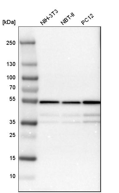
    Western Blot: ATPB Antibody - Western blot analysis in mouse cell line NIH-3T3, rat cell line NBT-II and rat cell line pC12.
    IHC Image

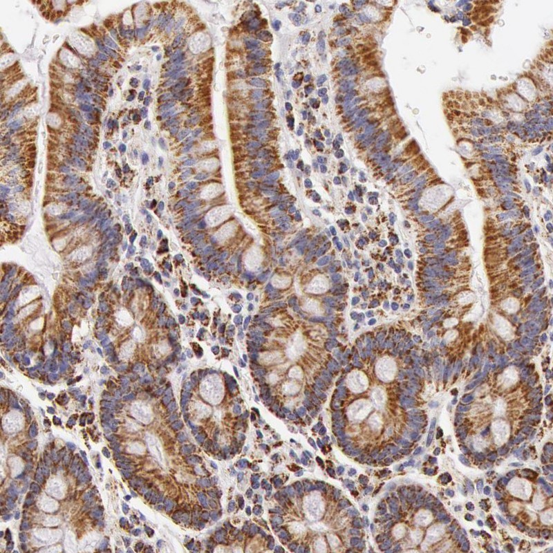
    Immunohistochemistry-Paraffin: ATPB Antibody - Staining of human duodenum shows moderate granular cytoplasmic positivity in glandular cells.
    ICC/IF Image

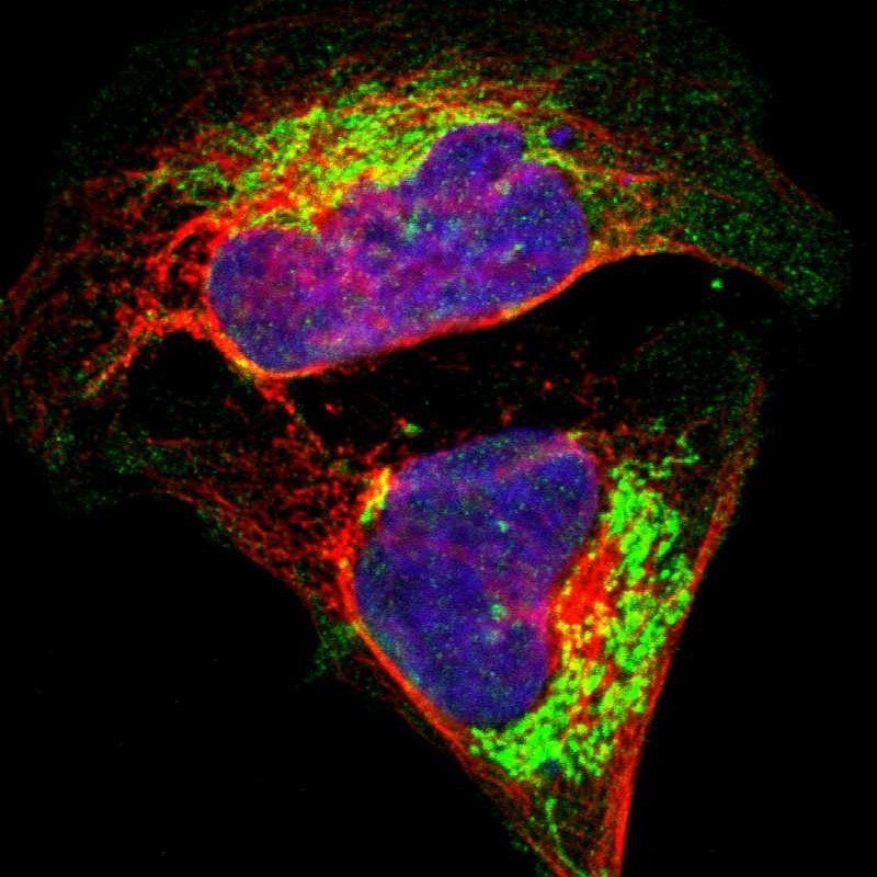
    Immunocytochemistry/Immunofluorescence: ATPB Antibody - Immunofluorescent staining of human cell line U-2 OS shows localization to mitochondria.

    Related Products

    Reviews

    loading...
    Content *:
    Customer Review :
    Verification Code * :
    refresh image

    Shipping and Paying Info
    ......
    Order Information
    You can place an order online step by step as blow.

    Step 1. Register for a new account and Log in, Find out your product by searching our website.
    Step 2. Add the Product into your shopping basket, select your settlement currency.
    Step 3. Checkout by Paypal, Pay by credit card or bank transfer for your order.
    Step 4. Ship the goods for you, providing you package tracking numbers. 

    Any questions, please contact us via email: sale@engibodybio.com
    Payments by
    All our products are For Research Use Only. Not for diagnostic or therapeutic usages.