CoIP, ChIP, CUT&RUN, CUT&Tag Expert! My Basket (...)
Worldwide delivery!
Having the Best Alpaca Nanobody Products!
Home | About us | Contact us | How to order |
    My Base Info
    Shopping Basket
    My Orders
    My Shipping Info
    Modify Password
    Log Out

    Anti-Bak Rabbit mAb

    Catalog number :AT0777
    loading...
    Background: 
    Bak is a proapoptotic member of the Bcl-2 family. This protein is located on the outer membrane of mitochondria and is an essential component for transduction of apoptotic signals through the mitochondrial pathway. Upon apoptotic stimulation, an upstream stimulator like truncated BID (tBID) induces conformational changes in Bak to form oligomer channels in the mitochondrial membrane for cytochrome c release. The release of cytochrome c to the cytosol activates the caspase-9 pathway and eventually leads to cell death.
     
    UniProt ID: Q16611
    Overview
    Reactivity
    Human, Mouse, Rat, Bovine, Pig, Monkey
    Tested applications
    Western Blotting: 1/1000-1/2000
    Immunofluorescence: 1/500
    Immunohistochemistry (Paraffin): 1/500
    Immunoprecipitation(IP) : 1/100 
    Optimal dilutions/concentrations should be determined by the end user. 
    Specificity
    This antibody detects endogenous levels of total Bak protein.
    Properties
    Immunogen
    a synthetic peptide corresponding to residues surrounding Gly75 of human Bak protein.
    Clonality
    Monoclonal, clone number: 1A4
    Isotype
    Rabbit IgG
    Form
    Liquid, 100 μl,1mg/ml, PBS (pH 7.2) and 40% Glycerol,0.02% Sodium Azide
    Storage instruction
    Store at +4°C short term (1-2 weeks). Aliquot and store at -20°C, Avoid freeze / thaw cycle.
    Host
    Rabbit
    Applications
    WB Image

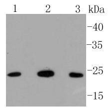
    WB: BAK Antibody - Analysis of Bak on different lysates using anti-Bak antibody at 1/1,000 dilution. Positive control: Lane 1: Hela Lane 2: Human skeletal muscle Lane 3: Ags
    IHC Image

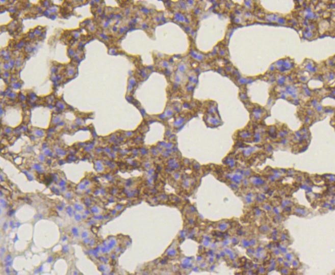
    Immunohistochemistry-Paraffin: BAK Antibody - Analysis of paraffin-embedded mouse lung tissue using anti-Bak antibody. Counter stained with hematoxylin.
    ICC/IF Image

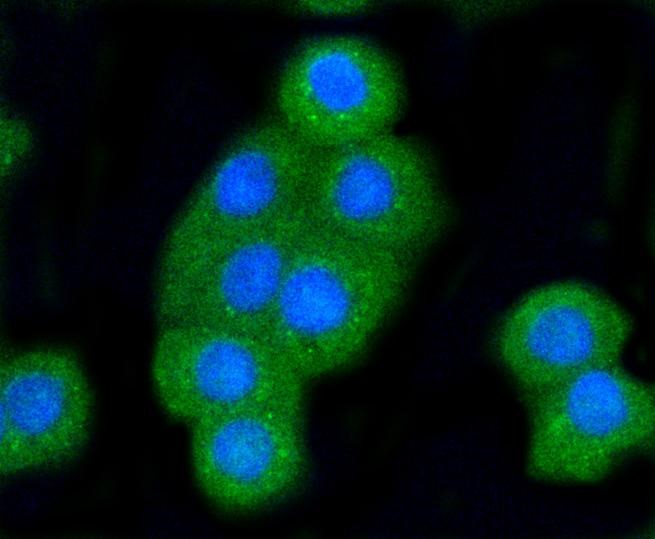
    Immunocytochemistry/Immunofluorescence: BAK Antibody - Staining Bak in CRC cells (green). The nuclear counter stain is DAPI (blue). Cells were fixed in paraformaldehyde, permeabilised with 0.25% Triton X100/PBS.

    Related Products

    Reviews

    loading...
    Content *:
    Customer Review :
    Verification Code * :
    refresh image

    Shipping and Paying Info
    ......
    Order Information
    You can place an order online step by step as blow.

    Step 1. Register for a new account and Log in, Find out your product by searching our website.
    Step 2. Add the Product into your shopping basket, select your settlement currency.
    Step 3. Checkout by Paypal, Pay by credit card or bank transfer for your order.
    Step 4. Ship the goods for you, providing you package tracking numbers. 

    Any questions, please contact us via email: sale@engibodybio.com
    Payments by
    All our products are For Research Use Only. Not for diagnostic or therapeutic usages.