Anti-Bcl-2 Rabbit mAb
Catalog number :AT0377
Bcl-2 exerts a survival function in response to a wide range of apoptotic stimuli through inhibition of mitochondrial cytochrome c release. It has been implicated in modulating mitochondrial calcium homeostasis and proton flux. Several phosphorylation sites have been identified within Bcl-2 including Thr56, Ser70, Thr74 and Ser87. It has been suggested that these phosphorylation sites may be targets of the ASK1/MKK7/JNK1 pathway, and that phosphorylation of Bcl-2 may be a marker for mitotic events. Mutation of Bcl-2 at Thr56 or Ser87 inhibits its anti-apoptotic activity during glucocorticoid-induced apoptosis of T lymphocytes. Interleukin 3 and JNK-induced Bcl-2 phosphorylation at Ser70 may be required for its enhanced anti-apoptotic functions.
- Overview
- Description
- Rabbit monoclonal antibody to Bcl2
- Reactivity
- Reacts with: Mouse, Human, Rat
- Tested applications
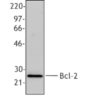
IHC-P : Use at an assay dependent concentration. ICC/IF : Use at an assay dependent concentration. WB : Use a concentration of 1/500. Detects a band of approximately 26 kDa (predicted molecular weight: 26 kDa).
- Properties
- Immunogen
- Synthetic peptide conjugated to KLH derived from within residues 1 - 100 of Human Bcl2.
- Clonality
- Monoclonal, clone number: 4B7
- Isotype
- Rabbit IgG
- Form
- Liquid
Preservative: 0.02% Sodium Azide
Constituents: 1% BSA, PBS, pH 7.4Liquid
- Storage instruction
- Store at +4°C short term (1-2 weeks). Aliquot and store at -20°C or -80°C. Avoid repeated freeze / thaw cycles.
- Database links
- Entrez Gene: 403416 Dog
- Entrez Gene: 596 Human
- Entrez Gene: 12043 Mouse
- Entrez Gene: 24224 Rat
- Omim: 151430 Human
- SwissProt: P10415 Human
- SwissProt: P10417 Mouse
- SwissProt: P49950 Rat
- Applications
- WB Image
 Anti-Bcl2 antibody (AT0377) at 1/500 + mouse splenocytes Whole Cell Lysate at 20 µg
Secondary
Goat polyclonal Secondary Antibody to Rabbit IgG - H&L (HRP) at 1/3000 dilution
developed using the ECL technique
Performed under reducing conditions.
Predicted band size : 26 kDa
Observed band size : 26 kDa
- ICC/IF Image
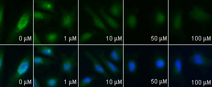
 AT0377 staining Bcl2 in HeLa cells treated with bisdemethoxycurcumin, by ICC/IF. Decrease of Bcl2 expression correlates with increased concentration of bisdemethoxycurcumin, as described in literature.
Immunocytochemistry/ Immunofluorescence - Anti-Bcl2 antibody
AT0377 stained HepG2 cells. The cells were 4% formaldehyde fixed (10 min) and then incubated in 1%BSA / 10% normal goat serum / 0.3M glycine in 0.1% PBS-Tween for 1h to permeabilise the cells and block non-specific protein-protein interactions. The cells were then incubated with the antibody AT0377 at 5µg/ml overnight at +4°C. The secondary antibody (green) was DyLight® 488 goat anti- rabbit IgG (H+L) used at a 1/1000 dilution for 1h. Alexa Fluor® 594 WGA was used to label plasma membranes (red) at a 1/200 dilution for 1h. DAPI was used to stain the cell nuclei (blue) at a concentration of 1.43µM.