Anti-CNPase Rabbit mAb - Oligodendrocyte Marker
Catalog number :AT0680
CNPase (2', 3’-cyclic nucleotide 3'-phosphodiesterase) catalyzes the in vitro hydrolysis of 2’, 3’-cyclic nucleotides to produce 2’-nucleotides. The in vivo molecular function and native substrate of this nucleotide phosphodiesterase remains under investigation. High CNPase expression is seen in oligodendrocytes and Schwann cells as CNPase accounts for roughly 4% of the total myelin protein in the central nervous system. CNPase binds to tubulin heterodimers and plays a role in tubulin polymerization, and oligodendrocyte process outgrowth. Typical myelination is seen in CNPase knock-out mice, but they suffer severe neurodegeneration from axonal loss and oligodendrocytes display abnormal paranodal loop structure prior to axonal degeneration. Paranodal loops typically contact the axolemma in axon-glial signaling; neurodegeneration in CNPase knock-out mice is a secondary consequence of impaired cell-cell communication.
- Overview
- Reactivity
- Human, Mouse, Rat, Monkey
- Tested applications
- Western Blotting 1:1000Immunoprecipitation 1:200Immunohistochemistry (Paraffin) 1:500Optimal dilutions/concentrations should be determined by the end user.
- Specificity
- This antibody detects endogenous levels of total CNPase protein.
- Properties
- Immunogen
- Synthetic peptide conjugated to KLH derived from within residues 350 to the C-terminus of Human CNPase.
- Clonality
- Monoclonal, clone number: 4S2
- Isotype
- Rabbit IgG
- Form
- Liquid, 100 μl,1mg/ml, PBS (pH 7.2) and 40% Glycerol,0.02% Sodium Azide
- Storage instruction
- Store at +4°C short term (1-2 weeks). Aliquot and store at -20°C, Avoid freeze / thaw cycle.
- Host
- Rabbit
- Applications
- WB Image

Western Blot: CNPase Antibody - Analysis of CNPase on mouse brain lysates using anti-CNPase antibody at 1/1,000 dilution.
- IHC Image

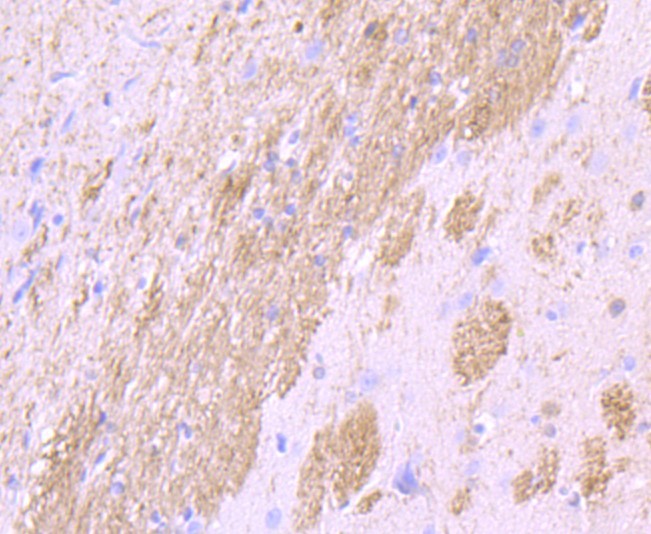
Immunohistochemistry-Paraffin: CNPase Antibody - Analysis of paraffin-embedded rat brain tissue using anti-CNPase antibody. Counter stained with hematoxylin.
- ICC/IF Image

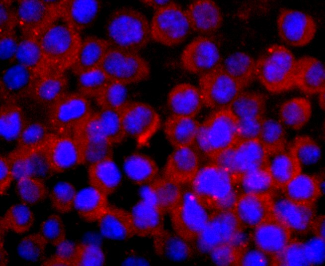
Immunocytochemistry/Immunofluorescence: CNPase Antibody- Staining CNPase in N2A cells (green). Cells were fixed in paraformaldehyde, permeabilised with 0.25% Triton X100/PBS.
Related Products
Reviews
loading...