Protein tyrosine-kinase transmembrane receptor for CSF1 and IL34.
Anti-CSF1R Rabbit mAb
Catalog number :AT0418
- Overview
- Description
- Rabbit monoclonal antibody to CSF1R
- Reactivity
- Human, Mouse, Rat
- Tested applications
- WB: 1/500 - 1/1000. Predicted molecular weight: 108 kDa.
IHC-P: 1/50 - 1/100.
ICC/IF: 1/500 - 1/1000.
- Properties
- Immunogen
- Synthesized non phosphopeptide derived from human MCSF Receptor around the phosphorylation site of tyrosine 809 (S-N-YP-I-V).
- Clonality
- Monoclonal, clone number: 5A5
- Isotype
- IgG
- Form
- Liquid
Preservative: 0.02% Sodium Azide
Constituents: 50% Glycerol, PBS (without Mg2+ and Ca2+), 150mM Sodium chloride, pH 7.4
- Storage instruction
- Shipped at 4°C. Upon delivery aliquot and store at -20°C. Avoid freeze / thaw cycles.
- Database links
- Entrez Gene: 1436 Human
- Entrez Gene: 12978 Mouse
- Omim: 164770 Human
- SwissProt: P07333 Human
- SwissProt: P09581 Mouse
- Unigene: 586219 Human
- Unigene: 22574 Mouse
- Applications
- WB Image
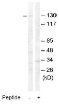
Western blot - MCSF Receptor antibody
All lanes : Anti-MCSF Receptor antibody (AT0418) at 1/500 dilution
Lane 1 : Extracts from HT-29 cells
Lane 2 : Extracts from HT-29 cells, plus peptide at 10 µg
Lysates/proteins at 5 µg per lane.
Predicted band size : 108 kDa
Observed band size : 130 kDa
Why is the actual band size different from the predicted?
Western blotting is a technique that separates proteins based on size - in general, the smaller the protein the faster it migrates through the gel. However, migration is also affected by other factors and so the actual band size observed may differ from that predicted. Common factors include...
- post-translational modification - e.g. phosphorylation, glycosylation etc which increases the size of the protein
- post-translation cleavage - e.g. many proteins are synthesized as pro-proteins, and then cleaved to give the active form, e.g. pro-caspases
- splice variants - alternative splicing may create different sized proteins from the same gene
- relative charge - the composition of amino acids (charged vs non-charged)
- multimers - e.g. dimerisation of a protein. This is usually prevented in reducing conditions, although strong interactions can result in the appearance of higher bands
- IHC Image
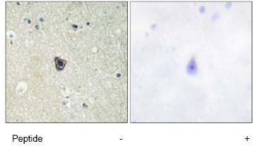
Immunohistochemistry (Formalin/PFA-fixed paraffin-embedded sections) - MCSF Receptor antibody
AT0418, at a 1/50 dilution, staining MCSF Receptor in paraffin embedded human brain tissue by Immunohistochemistry, in the absence (left image) or presence (right image) of the immunising peptide.
- ICC/IF Image

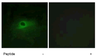
Immunocytochemistry/ Immunofluorescence - MCSF Receptor antibody
AT0418, at a 1/500 dilution, staining MCSF Receptor in HeLa cells by Immunofluorescence analysis in the absence (left image) or presence (right image) of the immunising peptide.
Related Products
Reviews
loading...

