CoIP, ChIP, CUT&RUN, CUT&Tag Expert! My Basket (...)
Worldwide delivery!
Having the Best Alpaca Nanobody Products!
Home | About us | Contact us | How to order |
    My Base Info
    Shopping Basket
    My Orders
    My Shipping Info
    Modify Password
    Log Out

    Anti-Caspase 3 (active/cleaved) Rabbit mAb

    Catalog number :AT0770
    loading...
    Cleavage by granzyme B, caspase-6, caspase-8 and caspase-10 generates the two active subunits. Additional processing of the propeptides is likely due to the autocatalytic activity of the activated protease. Active heterodimers between the small subunit of caspase-7 protease and the large subunit of caspase-3 also occur and vice versa.
    S-nitrosylated on its catalytic site cysteine in unstimulated human cell lines and denitrosylated upon activation of the Fas apoptotic pathway, associated with an increase in intracellular caspase activity. Fas therefore activates caspase-3 not only by inducing the cleavage of the caspase zymogen to its active subunits, but also by stimulating the denitrosylation of its active site thiol.
    Overview
    Description
    anti Caspase 3 Rabbit monoclonal antibody
    Reactivity
     Mouse, Rat, Rabbit, Human, Monkey, Pig
    Properties
    Immunogen
    Recombinant full length protein corresponding to Human Caspase-3.
    Database link: P42574
    Clonality
    Polyclonal
    Isotype
    Monoclonal, clone number: 2B7
    Form
    Liquid
    Storage instruction
    Shipped at 4°C. Store at +4°C short term (1-2 weeks). Store at -20°C or -80°C. Avoid freeze / thaw cycle.
    Applications
    WB Image

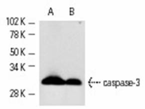
    Western blot analysis of caspase-3 precursor expression in HuT 78 (A) and CCRF-HSB-2 (B) whole cell lysates.
    ICC/IF Image

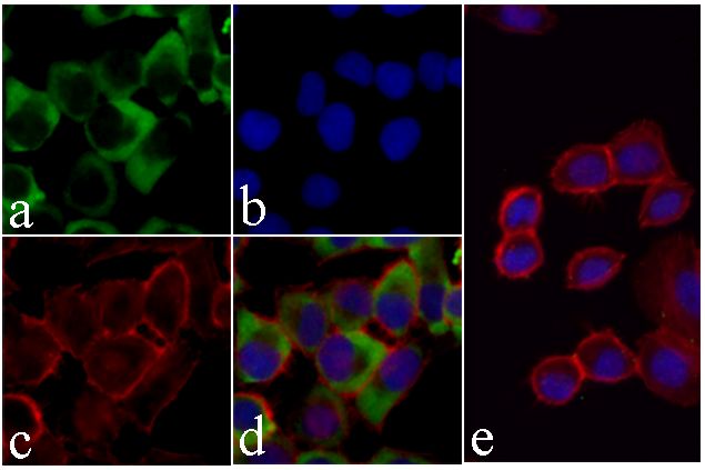
    Caspase 3 Antibody in IF
    Immunofluorescence analysis of Caspase 3 was done on 70% confluent log phase HeLa cells. The cells were fixed with 4% paraformaldehyde for 15 minutes, permeabilized with 0.25% Triton X-100 for 10 minutes, and blocked with 5% BSA for 1 hour at room temperature. The cells were labeled with Caspase-3 Antibody at a dilution of 1:500 in 1% BSA and incubated for 3 hours at room temperature and then labeled with Alexa Fluor 488 Goat Anti Rabbit IgG Secondary Antibody at a dilution of 1:400 for 30 minutes at room temperature (Panel a: green). Nuclei (Panel b: blue) were stained with SlowFade Gold Antifade Mountant with DAPI. F-actin (Panel c: red) was stained with Alexa Fluor 594 Phalloidin Panels d is a merged image showing cytoplasmic localization and panel e a is no primary antibody control. The images were captured using a Nikon microscope at 20X magnification.

    Related Products

    Reviews

    loading...
    Content *:
    Customer Review :
    Verification Code * :
    refresh image

    Shipping and Paying Info
    ......
    Order Information
    You can place an order online step by step as blow.

    Step 1. Register for a new account and Log in, Find out your product by searching our website.
    Step 2. Add the Product into your shopping basket, select your settlement currency.
    Step 3. Checkout by Paypal, Pay by credit card or bank transfer for your order.
    Step 4. Ship the goods for you, providing you package tracking numbers. 

    Any questions, please contact us via email: sale@engibodybio.com
    Payments by
    All our products are For Research Use Only. Not for diagnostic or therapeutic usages.