CoIP, ChIP, CUT&RUN, CUT&Tag Expert! My Basket (...)
Worldwide delivery!
Having the Best Alpaca Nanobody Products!
Home | About us | Contact us | How to order |
    My Base Info
    Shopping Basket
    My Orders
    My Shipping Info
    Modify Password
    Log Out

    Anti-E Cadherin Rabbit mAb - Intercellular Junction Marker

    Catalog number :AT0800
    loading...
    Background: 
    Cadherins are a superfamily of transmembrane glycoproteins that contain cadherin repeats of approximately 100 residues in their extracellular domain. Cadherins mediate calcium-dependent cell-cell adhesion and play critical roles in normal tissue development. The classic cadherin subfamily includes N-, P-, R-, B-, and E-cadherins, as well as about ten other members that are found in adherens junctions, a cellular structure near the apical surface of polarized epithelial cells. The cytoplasmic domain of classical cadherins interacts with β-catenin, γ-catenin (also called plakoglobin), and p120 catenin. β-catenin and γ-catenin associate with α-catenin, which links the cadherin-catenin complex to the actin cytoskeleton. While β- and γ-catenin play structural roles in the junctional complex, p120 regulates cadherin adhesive activity and trafficking. Investigators consider E-cadherin an active suppressor of invasion and growth of many epithelial cancers. Research studies indicate that cancer cells have upregulated N-cadherin in addition to loss of E-cadherin. This change in cadherin expression is called the "cadherin switch." N-cadherin cooperates with the FGF receptor, leading to overexpression of MMP-9 and cellular invasion. Research studies have shown that in endothelial cells, VE-cadherin signaling, expression, and localization correlate with vascular permeability and tumor angiogenesis. Investigators have also demonstrated that expression of P-cadherin, which is normally present in epithelial cells, is also altered in ovarian and other human cancers.
     
    UniProt ID: 
    P12830
     
    Overview
    Reactivity
    Human, Mouse, Rat, Bovine, Pig, Monkey
    Tested applications
    Western Blotting: 1/1000-1/2000
    Immunofluorescence: 1/500
    Immunohistochemistry (Paraffin): 1/500
    Immunoprecipitation(IP) : 1/100 
    Optimal dilutions/concentrations should be determined by the end user. 
    Specificity
    This antibody detects endogenous levels of total E-cadherin protein. This antibody does not cross-react with other cadherin proteins.
    Properties
    Immunogen
    Recombinant protein encompassing a sequence within the center region of human E-Cadherin. The exact sequence is proprietary.
    Clonality
    Monoclonal, clone number: 6B1
    Isotype
    Rabbit IgG
    Form
    Liquid, 100 μl,1mg/ml, PBS (pH 7.2) and 40% Glycerol,0.02% Sodium Azide
    Storage instruction
    Store at +4°C short term (1-2 weeks). Aliquot and store at -20°C, Avoid freeze / thaw cycle.
    Host
    Rabbit
    Applications
    WB Image

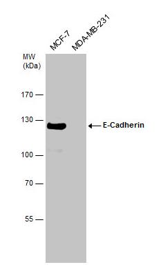
    Genetic Strategies Validation. Western Blot: E-Cadherin Antibody - Non-transfected (-) and transfected (+) MCF-7 whole cell extracts (30 ug) were separated by 5% SDS-PAGE, and the membrane was blotted with E-Cadherin antibody.
    IHC Image

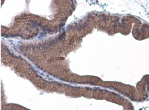
    Immunohistochemistry-Paraffin: E-Cadherin Antibody - Paraffin-embedded rat prostate. E-Cadherin antibody diluted at 1:500.
    ICC/IF Image

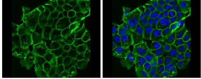
    Immunocytochemistry/Immunofluorescence: E-Cadherin Antibody - A431 cells were fixed in 4% paraformaldehyde at RT for 15 min. Green: E-Cadherin protein stained by E-Cadherin antibody diluted at 1:500. Blue: Hoechst 33342 staining.

    Related Products

    Reviews

    loading...
    Content *:
    Customer Review :
    Verification Code * :
    refresh image

    Shipping and Paying Info
    ......
    Order Information
    You can place an order online step by step as blow.

    Step 1. Register for a new account and Log in, Find out your product by searching our website.
    Step 2. Add the Product into your shopping basket, select your settlement currency.
    Step 3. Checkout by Paypal, Pay by credit card or bank transfer for your order.
    Step 4. Ship the goods for you, providing you package tracking numbers. 

    Any questions, please contact us via email: sale@engibodybio.com
    Payments by
    All our products are For Research Use Only. Not for diagnostic or therapeutic usages.