Receptor tyrosine kinase binding ligands of the EGF family and activating several signaling cascades to convert extracellular cues into appropriate cellular responses. Known ligands include EGF, TGFA/TGF-alpha, amphiregulin, epigen/EPGN, BTC/betacellulin, epiregulin/EREG and HBEGF/heparin-binding EGF. Ligand binding triggers receptor homo- and/or heterodimerization and autophosphorylation on key cytoplasmic residues. The phosphorylated receptor recruits adapter proteins like GRB2 which in turn activates complex downstream signaling cascades. Activates at least 4 major downstream signaling cascades including the RAS-RAF-MEK-ERK, PI3 kinase-AKT, PLCgamma-PKC and STATs modules. May also activate the NF-kappa-B signaling cascade. Also directly phosphorylates other proteins like RGS16, activating its GTPase activity and probably coupling the EGF receptor signaling to the G protein-coupled receptor signaling. Also phosphorylates MUC1 and increases its interaction with SRC and CTNNB1/beta-catenin.
Isoform 2 may act as an antagonist of EGF action.
Anti-EGFR Rabbit mAb
Catalog number :AT0356
- Overview
- Description
- Rabbit monoclonal antibody to EGFR
- Reactivity
- Mouse, Rat, Sheep, Cow, Dog, Human
- Tested applications
Flow Cyt : Use at an assay dependent dilution. ICC/IF : 1/20 - 1/100. ICC : Use at an assay dependent dilution. WB : 1/200. IP : Use at an assay dependent dilution. IHC-P : Use at an assay dependent dilution. IHC-Fr : Use at an assay dependent dilution. IHC-FrFl : Use at an assay dependent concentration. In-Cell ELISA : Use at an assay dependent concentration.
- Properties
- Immunogen
Synthetic peptide: EDMDDVVDADEY, corresponding to C terminal amino acids 1005-1016 of EGFR.
- Clonality
- Monoclonal, clone number: 5A31
- Isotype
- IgG
- Form
Liquid
Preservative: 0.1% Sodium Azide
Constituents: 0.2% Gelatin, PBS
- Storage instruction
- Store at +4°C short term (1-2 weeks). Store at -20°C or -80°C. Avoid freeze / thaw cycle.
 Run BLAST with
- Applications
- WB Image
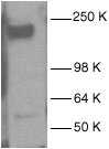 AT0356 at a 1/200 dilution staining A431 whole cell lysate.
- IHC Image
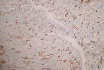
Immunohistochemistry (Formalin/PFA-fixed paraffin-embedded sections) - EGFR antibody
AT0356 staining formalin fixed paraffin-embedded sections of human adhesion tissue. The section was subjected to heat-mediated antigen retrieval in citrate buffer (pH 6.0) and blocked with 10% BSA for 1 hour prior to incubating with the primary antibody (diluted 1/200) for 12 hours at 4°C. A biotinylated goat anti-rabbit antibody was used as the secondary.
- ICC/IF Image
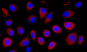 Cells were stained with anti-EGFR (AT0356; red) and DAPI (blue). A 1/40 dilution of AT0356 was used.HeLa cells were fixed with 4% formaldehyde in PEM buffer. The coverslip was incubated in blocking buffer of 5% powdered milk in TBS-T plus 0.02% sodium azide for 1 hour at room temperature. Blocking buffer was removed and primary antibody was added at a dilution of 1/40 and incubated overnight at 4 degrees celsius. The coverslips were then washed 4-5 times with blocking buffer for 5 minutes. Secondary antibody was added at a dilution of 1/1000 and incubated at room temperature for one hour. From this point on coverslips were covered with foil to protect them from light. They were washed 5 times with TBS-T and then one time with PEM, for 5 minutes each wash. The coverslips were fixed 10-30 minutes in 4% formaldehyde in PEM buffer, then washed 3 times with PEM buffer for 5 minutes. 0.1M ammonium chloride in PEM buffer was added for 10 minutes to quench auto-florescence, and then slips were washed 2 times for 5 minutes in PEM followed by 3 washes for 5 minutes in TBS-T. Coverslips were then counterstained with DAPI in TBS-T for 1-2 minutes, TBS-T was then added and the coverslips mounted.
- IP/CoIP Image
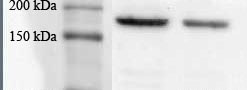 AT0356 immunoprecipitate of EGFR in human AGS whole cell lysate. 100 µg of cell lysate was incubated with primary antibody (1/100 in RIPA buffer) and matrix (Protein A/G) for 16 hours at 4°C.