Anti-GFP tag Rabbit pAb - for ChIP, CUT&RUN and CUT&Tag Grade
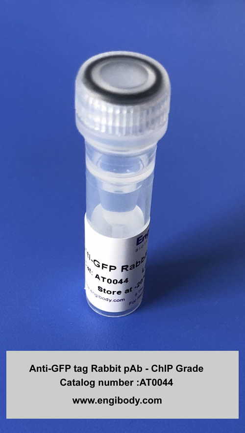
Catalog number :AT0044
Green fluorescence protein (GFP) is a 27 kDa protein derived from jellyfish Aequorea victoria. It emits green light (emission peak at a wavelength of 509 nm) when excited by blue light (excitation peak at a wavelength of 395 nm). GFP has become a very useful tool as a fusion protein to report gene expression, trace cell lineage and define subcellular protein localizations. YFP differs from GFP due to a mutation at T203Y.
- Overview
- Description
- Anti-GFP Rabbit polyclonal antibody, A mixture of 10 monoclonal antibodies for ChIP, CUT&RUN and CUT&Tag
- Reactivity
- Anti-GFP Antibody detects GFP-tagged proteins within cell lysates
- Tested applications
- WB: 1/1000-1/5000.
IHC/ICC/IF: 1/100-1/500.
Co-IP: 5µL per reaction
ChIP: 5-10µL per reaction
CUT&RUN: 1µL per reaction
CUT&Tag: 1µL per reaction
Optimal dilutions/concentrations should be determined by the end user.
- Product Picture

- Properties
- Immunogen
- Highly purified recombinant full length protein made in Escherichia coli. The antibody is directed against the entire GFP molecule.
- Clonality
- Polyclonal (A mixture of 10 monoclonal antibodies )
This is to be more suitable for capturing the different conformational epitopes of GFP fusion protein exposed in chromatin for ChIP, CUT&RUN and CUT&Tag
- Isotype
- Rabbit IgG
- Form
- Liquid
- Storage instruction
- Store at -20°C, Avoid freeze / thaw cycle. Do not aliquot the antibody.
- Applications
- WB Image

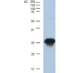
Anti-GFP antibody (AT0044) at 1/2000 dilution + Lysate prepared from rabbit reticulocytes at 30 µgDeveloped using the ECL techniquePerformed under reducing conditions.Observed band size: 27 kDaExposure time: 5 seconds
Recommended secondary antibody: Goat anti rabbit IgG-HRP (Engibody, Catalog number:AT0097)Recommended ECL: ECL Pico-Detect™ Western Chemiluminescent HRP Substrate (Engibody, Catalog number: IF6747)
- IHC Image

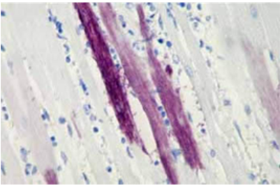
AT0044 staining dog hearts (Adv-GFP injection) tissue sections by IHC-P. Sections were PFA fixed and subjected to heat mediated antigen retrieval in citric acid (Ph6.0, 0.05% Tween20) prior to blocking with 10% serum for 30 mins at 37°C. The primary antibody was diluted 1/500 in PBS and incubated with the sample for 1 hour at 25°C.
- ICC/IF Image

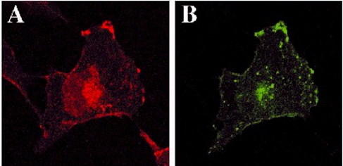
Immunofluorescence images showing similar localization of Yes-GFP (first 10 aa's of Yes PTK fused to the N-terminus of GFP) to full length Yes PTK.A: Distribution of Yes detected using mouse anti-Yes Ab followed by Texas Red-conjugated anti-mouse pAb.B: Chimeric GFP's detected using rabbit anti-GFP rabbit pAb (CAT: AT0044) followed by FITC-conjugated anti-rabbit pAb.
- Application Image

Related Products
Reviews
loading...