Anti-GSK3 beta Rabbit mAb
Catalog number :AT0350
Participates in the Wnt signaling pathway. Implicated in the hormonal control of several regulatory proteins including glycogen synthase, MYB and the transcription factor JUN. Phosphorylates JUN at sites proximal to its DNA-binding domain, thereby reducing its affinity for DNA. Phosphorylates MUC1 in breast cancer cells, and decreases the interaction of MUC1 with CTNNB1/beta-catenin. Phosphorylates CTNNB1/beta-catenin. Phosphorylates SNAI1. Plays an important role in ERBB2-dependent stabilization of microtubules at the cell cortex. Prevents the phosphorylation of APC and CLASP2, allowing its association with the cell membrane. In turn, membrane-bound APC allows the localization of MACF1 to the cell membrane, which is required for microtubule capture and stabilization. Phosphorylates MACF1 and this phosphorylation inhibits the binding of MACF1 to microtubules which is critical for its role in bulge stem cell migration and skin wound repair.
- Overview
- Description
- Rabbit monoclonal antibody to GSK3 beta
- Reactivity
- Reacts with: Mouse, Rat, Human
Predicted to work with: Rabbit, Horse, Chicken, Cow, Dog, Pig, Chimpanzee, Zebrafish, Macaque Monkey, Gorilla, Chinese Hamster
- Tested applications
IHC-P : Use a concentration of 1 µg/ml. Perform heat mediated antigen retrieval with Tris/EDTA buffer pH 9.0 before commencing with IHC staining protocol. WB : Use a concentration of 1 µg/ml. Detects a band of approximately 46 kDa (predicted molecular weight: 46 kDa).
- Properties
- Immunogen
- Synthetic peptide conjugated to KLH derived from within residues 300 - 400 of Mouse GSK3 beta.
- Clonality
- Monoclonal, clone number: 4TA7
- Isotype
- IgG
- Form
- pH: 7.40
Preservative: 0.02% Sodium azide
Constituent: PBS
Note: Batches of this product that have a concentration < 1mg/ml may have BSA added as a stabilising agent. If you would like information about the formulation of a specific lot, please contact our scientific support team who will be happy to help.Liquid
- Storage instruction
- Store at +4°C short term (1-2 weeks). Aliquot and store at -20°C or -80°C. Avoid repeated freeze / thaw cycles.
- Applications
- WB Image
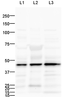 All lanes : Anti-GSK3 beta antibody (AT0350) at 1 µg/ml
Lane 1 : Heart (Mouse) Tissue Lysate
Lane 2 :Heart (Human) Tissue Lysate - adult normal tissue (AT0350)
Lane 3 : Heart (Rat) Tissue Lysate
Lysates/proteins at 25 µg per lane.
Secondary
Goat polyclonal Secondary Antibody to Rabbit IgG - H&L (HRP) at 1/10000 dilution
developed using the ECL technique
Performed under reducing conditions.
Predicted band size : 46 kDa
Observed band size : 46 kDa
Additional bands at : 23 kDa,52 kDa,73 kDa. We are unsure as to the identity of these extra bands.
Exposure time : 6 minutesThis blot was produced using a 10% Bis-tris gel under the MOPS buffer system. The gel was run at 200V for 50 minutes before being transferred onto a Nitrocellulose membrane at 30V for 70 minutes. The membrane was then blocked for an hour using 5% Bovine Serum Albumin before being incubated with ab124661 overnight at 4°C. Antibody binding was detected using an anti-rabbit antibody conjugated to HRP, and visualised using ECL development solution.
- IHC Image
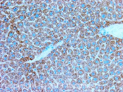 Immunohistochemistry (Formalin/PFA-fixed paraffin-embedded sections) - Anti-GSK3 beta antibody (AT0350)
IHC image of GSK3 beta staining in Mouse normal liver formalin fixed paraffin embedded tissue section, performed on a Leica Bond™ system using the standard protocol B. The section was pre-treated using heat mediated antigen retrieval (EDTA based pH 9.0 solution) for 20 mins. The section was then incubated with AT0350, 1µg/ml, for 15 mins at room temperature. A Goat anti-Rabbit biotinylated secondary antibody was used to detect the primary, and visualized using an HRP conjugated ABC system. DAB was used as the chromogen. The section was then counterstained with haematoxylin and mounted with DPX.
For other IHC staining systems (automated and non-automated) customers should optimize variable parameters such as antigen retrieval conditions, primary antibody concentration and antibody incubation times.