Anti-HDAC1 antibody - ChIP, CUT&RUN and CUT&Tag Grade
Catalog number :AT0360
Responsible for the deacetylation of lysine residues on the N-terminal part of the core histones (H2A, H2B, H3 and H4). Histone deacetylation gives a tag for epigenetic repression and plays an important role in transcriptional regulation, cell cycle progression and developmental events. Histone deacetylases act via the formation of large multiprotein complexes. Deacetylates SP proteins, SP1 and SP3, and regulates their function. Component of the BRG1-RB1-HDAC1 complex, which negatively regulates the CREST-mediated transcription in resting neurons. Upon calcium stimulation, HDAC1 is released from the complex and CREBBP is recruited, which facilitates transcriptional activation. Deacetylates TSHZ3 and regulates its transcriptional repressor activity. Deacetylates 'Lys-310' in RELA and thereby inhibits the transcriptional activity of NF-kappa-B.
- Overview
- Description
- Rabbit monoclonal antibody to HDAC1
- Reactivity
- Rat, Human, human, Xenopus laevis, Zebrafish
- Tested applications
ChIP, CUT&RUN and CUT&TagIHC : 1/300. WB : 1/1000-1/5000 ICC/IF : 1/300-1/500
- Properties
- Immunogen
- Synthetic peptide conjugated to KLH derived from within residues 50 - 150 of human HDAC1.
- Clonality
- Monoclonal
- Isotype
- Rabbit IgG
- Form
- Liquid
Preservative: 0.02% Sodium Azide
Constituents: 1% BSA, PBS, pH 7.4
- Storage instruction
- Store at +4°C short term (1-2 weeks). Aliquot and store at -20°C or -80°C. Avoid repeated freeze / thaw cycles.
- Applications
- WB Image

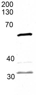
Anti-HDAC1 antibody (AT0360) at 1/3000 + whole embryo zebrafish tissue at 45 µg
Predicted band size : 56 kDa
Observed band size : 56 kDa
Additional bands at : 32 kDa (possible cross reactivity),37 kDa (possible cross reactivity).
- ICC/IF Image

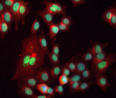
ICC/IF image of AT0360 stained human MCF7 cells. The cells were methanol fixed (5 min), permabilised in 0.1% PBS-Tween (20 min) and incubated with the antibody (AT0360, 1/300) for 1h at room temperature. 1%BSA / 10% normal goat serum / 0.3M glycine was used to block non-specific protein-protein interactions. The secondary antibody (green) was Alexa Fluor® 488 goat anti-rabbit IgG (H+L) used at a 1/1000 dilution for 1h. Alexa Fluor® 594 WGA was used to label plasma membranes (red). DAPI was used to stain the cell nuclei (blue). This antibody also gave a positive IF result in HeLa, HEK 293 and HepG2 cells.
Related Products
Reviews
loading...