Functions as a master transcriptional regulator of the adaptive response to hypoxia. Under hypoxic conditions activates the transcription of over 40 genes, including, erythropoietin, glucose transporters, glycolytic enzymes, vascular endothelial growth factor, and other genes whose protein products increase oxygen delivery or facilitate metabolic adaptation to hypoxia. Plays an essential role in embryonic vascularization, tumor angiogenesis and pathophysiology of ischemic disease. Binds to core DNA sequence 5'-[AG]CGTG-3' within the hypoxia response element (HRE) of target gene promoters. Activation requires recruitment of transcriptional coactivators such as CREBPB and EP300. Activity is enhanced by interaction with both, NCOA1 or NCOA2. Interaction with redox regulatory protein APEX seems to activate CTAD and potentiates activation by NCOA1 and CREBBP.
Anti-HIF1 alpha Rabbit mAb
Catalog number :AT0412
- Overview
- Description
- Rabbit monoclonal antibody to HIF1 alpha
- Reactivity
- Human, Mouse, Rat
- Tested applications
- WB: 1/500 - 1/1000.
IHC-P: 1/50 - 1/500
ICC:1/50 - 1/200
CoIP:1/50 - 1/200
- Properties
- Immunogen
Synthetic peptide corresponding to a region within N terminal amino acids 1-31 of Human HIF1 alpha, conjugated to KLH.
- Clonality
- Monoclonal, clone number: 6A6
- Isotype
- IgG
- Form
- Liquid
Preservative: 0.09% Sodium Azide
Constituents: PBS
- Storage instruction
- Store at 4°C (up to 6 months). For long term storage store at -20°C
- Database links
- Entrez Gene: 281814 Cow
- Entrez Gene: 3091 Human
- Omim: 603348 Human
- SwissProt: Q9XTA5 Cow
- SwissProt: Q16665 Human
- Unigene: 597216 Human
- Applications
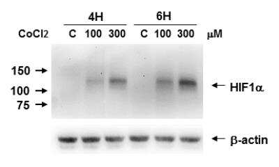
- WB Figure 1

Anti-HIF1 alpha antibody (AT0412) at 1/500 dilution + by CoCl2 on Caki-1 cell lysate at 35 µg
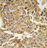
- IHC Image

AT0412, at a 1/50 dilution, staining HIF1 alpha in formalin fixed and paraffin embedded lung carcinoma by by Immunohistochemistry, followed by peroxidase conjugation of the secondary antibody and DAB staining.
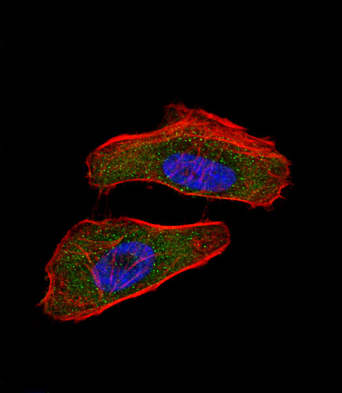
- ICC/IF Image

Immunofluorescence of HeLa cells labelling HIF1 alpha with AT0412. Hela cells were fixed with 4% PFA (20 min), permeabilized with Triton X-100 (0.1%, 10 min), then incubated with AT0412 (1:25, 1 h at 37℃). Alexa Fluor® 488 conjugated donkey anti-rabbit antibody (green) was used as the secondary antibody (1:400, 50 min at 37℃). Cytoplasmic actin was counterstained with Alexa Fluor® 555 (red) conjugated Phalloidin (7units/ml, 1 h at 37℃) and nuclei were counterstained with DAPI (blue) (10 µg/ml, 10 min). HIF1 alpha immunoreactivity is localized to the cytoplasm and nucleus.
Related Products
Reviews
loading...