CoIP, ChIP, CUT&RUN, CUT&Tag Expert! My Basket (...)
Worldwide delivery!
Having the Best Alpaca Nanobody Products!
Home | About us | Contact us | How to order |
    My Base Info
    Shopping Basket
    My Orders
    My Shipping Info
    Modify Password
    Log Out

    Anti-HSD3B2 (3 beta HSD) Rabbit mAb

    Catalog number :AT0816
    loading...
    3-beta-HSD is a bifunctional enzyme, that catalyzes the oxidative conversion of Delta(5)-ene-3-beta-hydroxy steroid, and the oxidative conversion of ketosteroids. The 3-beta-HSD enzymatic system plays a crucial role in the biosynthesis of all classes of hormonal steroids.
    Expressed in adrenal gland, testis and ovary.
    Defects in HSD3B2 are the cause of adrenal hyperplasia type 2 (AH2) [MIM:201810]. AH2 is a form of congenital adrenal hyperplasia, a common recessive disease due to defective synthesis of cortisol. Congenital adrenal hyperplasia is characterized by androgen excess leading to ambiguous genitalia in affected females, rapid somatic growth during childhood in both sexes with premature closure of the epiphyses and short adult stature. Four clinical types: 'salt wasting' (SW, the most severe type), 'simple virilizing' (SV, less severely affected patients), with normal aldosterone biosynthesis, 'non-classic form' or late onset (NC or LOAH), and 'cryptic' (asymptomatic). In AH2, virilization is much less marked or does not occur. AH2 is frequently lethal in early life.
    Overview
    Reactivity
    Mouse, Rat, Human
    Tested applications
    Western Blotting 1:1000
    IHC 1:50-1:500
    ICC 1:50-1:500
     
    Optimal dilutions/concentrations should be determined by the end user.
    Specificity
    This antibody recognizes endogenous levels of total HSD3B2 protein.
    Properties
    Immunogen
    Synthetic peptide corresponding to Human HSD3B2 N terminal.
    Clonality
    Monoclonal, clone number: 7F2
    Isotype
    Rabbit IgG
    Form
    Liquid, 100 μl,1mg/ml, PBS (pH 7.2) and 40% Glycerol,0.02% Sodium Azide
    Storage instruction
    Store at -20°C
    Host
    Rabbit
    Applications
    WB Image

    Anti-HSD3B2 antibody at 1 :1000 dilution
    + 293T cell lysate at 10 µg
    Secondary
    HRP conjugated anti-Rabbit IgG at 1:5000 dilution
    band size: 42 kDa
     
    ICC/IF Image

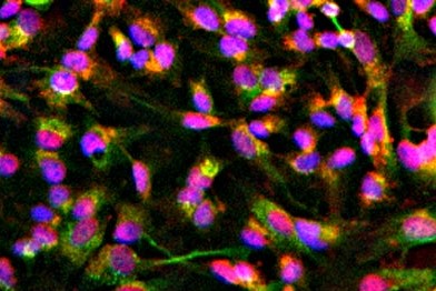
    ICC/IF image of AT0816 stained HepG2 cells. The cells were 100% methanol fixed (5 min) and then incubated in 1%BSA / 10% normal goat serum / 0.3M glycine in 0.1% PBS-Tween for 1h to permeabilise the cells and block non-specific protein-protein interactions. The cells were then incubated with the antibody (AT0816, 1:500) overnight at +4°C. The secondary antibody (green) was DyLight® 488 goat anti-rabbit IgG (H+L) used at a 1:1000 dilution for 1h. Alexa Fluor® 594 WGA was used to label plasma membranes (red) at a 1:200 dilution for 1h. DAPI was used to stain the cell nuclei (blue) at a concentration of 1.43µM.

    Related Products

    Reviews

    loading...
    Content *:
    Customer Review :
    Verification Code * :
    refresh image

    Shipping and Paying Info
    ......
    Order Information
    You can place an order online step by step as blow.

    Step 1. Register for a new account and Log in, Find out your product by searching our website.
    Step 2. Add the Product into your shopping basket, select your settlement currency.
    Step 3. Checkout by Paypal, Pay by credit card or bank transfer for your order.
    Step 4. Ship the goods for you, providing you package tracking numbers. 

    Any questions, please contact us via email: sale@engibodybio.com
    Payments by
    All our products are For Research Use Only. Not for diagnostic or therapeutic usages.