Histones H1 are necessary for the condensation of nucleosome chains into higher-order structures. The H1F0 histones are found in cells that are in terminal stages of differentiation or that have low rates of cell division.Belongs to the histone H1/H5 family.Contains 1 H15 (linker histone H1/H5 globular) domain.
Anti-Histone H1.0 Rabbit mAb - ChIP, CUT&RUN and CUT&Tag Grade
Catalog number :AT0713
- Overview
- Description
- Anti-Histone H1.0 Rabbit Monoclonal antibody
- Reactivity
- Human, mouse, rat, bovine, chicken, Drosophilia melanogaster
- Tested applications
- Western Blotting 1:1000
Immunofluorescence 1:500
Immunohistochemistry (Paraffin) 1:500
Immunoprecipitation(IP) 1;100
ChIP, CUT&RUN and CUT&Tag
Optimal dilutions/concentrations should be determined by the end user.
- Specificity
- This antibody detects endogenous levels of total Histone H1.0 protein.
- Properties
- Immunogen
- Recombinant fragment within Human Histone H1.0 aa 1-194. The exact sequence is proprietary.
- Clonality
- Monoclonal, clone number: 4B7
- Isotype
- Rabbit IgG
- Form
- Liquid, 100 μl,1mg/ml, PBS (pH 7.2) and 40% Glycerol,0.02% Sodium Azide
- Storage instruction
- Store at +4°C short term (1-2 weeks). Aliquot and store at -20°C, Avoid freeze / thaw cycle.
- Database links
- P07305
- Host
- Rabbit
- Applications
- WB Image
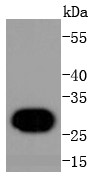
Western Blot: Histone H1.0 Antibody - Analysis of Histone H1.0 on human lung lysates using anti-Histone H1.0 antibody at 1/1,000 dilution.
- IHC Image
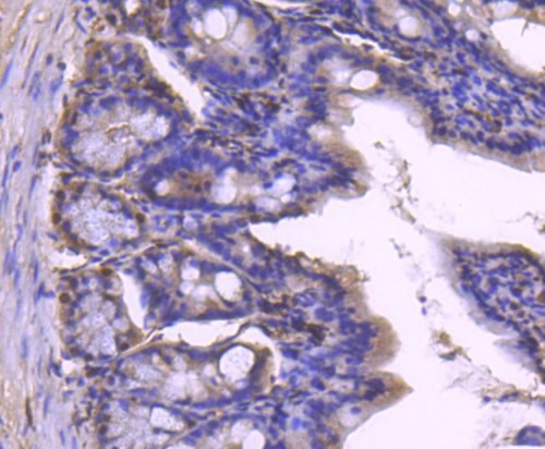
Immunohistochemistry-Paraffin: Histone H1.0 Antibody - Analysis of paraffin-embedded mouse colon tissue using anti-Histone H1.0 antibody. Counter stained with hematoxylin.
- ICC/IF Image
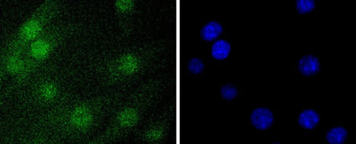
Immunocytochemistry/Immunofluorescence: Histone H1.0 Antibody - Staining Histone H1.0 in NIH/3T3 cells (green). The nuclear counter stain is DAPI (blue). Cells were fixed in paraformaldehyde, permeabilised with 0.25% Triton X100/PBS.
Related Products
Reviews
loading...