CoIP, ChIP, CUT&RUN, CUT&Tag Expert! My Basket (...)
Worldwide delivery!
Having the Best Alpaca Nanobody Products!
Home | About us | Contact us | How to order |
    My Base Info
    Shopping Basket
    My Orders
    My Shipping Info
    Modify Password
    Log Out

    Anti-Histone H2A.X (phospho S139) Rabbit mAb - ChIP, CUT&RUN and CUT&Tag Grade

    Catalog number :AT0729
    loading...
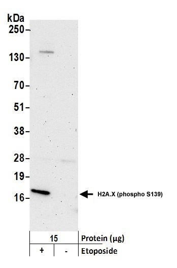
    Histone H2A.X is a variant histone that represents approximately 10% of the total H2A histone proteins in normal human fibroblasts (1). H2A.X is required for checkpoint-mediated cell cycle arrest and DNA repair following double-stranded DNA breaks (1). DNA damage, caused by ionizing radiation, UV-light, or radiomimetic agents, results in rapid phosphorylation of H2A.X at Ser139 by PI3K-like kinases, including ATM, ATR, and DNA-PK (2,3). Within minutes following DNA damage, H2A.X is phosphorylated at Ser139 at sites of DNA damage (4). This very early event in the DNA-damage response is required for recruitment of a multitude of DNA-damage response proteins, including MDC1, NBS1, RAD50, MRE11, 53BP1, and BRCA1 (1). In addition to its role in DNA-damage repair, H2A.X is required for DNA fragmentation during apoptosis and is phosphorylated by various kinases in response to apoptotic signals. H2A.X is phosphorylated at Ser139 by DNA-PK in response to cell death receptor activation, c-Jun N-terminal Kinase (JNK1) in response to UV-A irradiation, and p38 MAPK in response to serum starvation (5-8). H2A.X is constitutively phosphorylated on Tyr142 in undamaged cells by WSTF (Williams-Beuren syndrome transcription factor) (9,10). Upon DNA damage, and concurrent with phosphorylation of Ser139, Tyr142 is dephosphorylated at sites of DNA damage by recruited EYA1 and EYA3 phosphatases (9). While phosphorylation at Ser139 facilitates the recruitment of DNA repair proteins and apoptotic proteins to sites of DNA damage, phosphorylation at Tyr142 appears to determine which set of proteins are recruited. Phosphorylation of H2A.X at Tyr142 inhibits the recruitment of DNA repair proteins and promotes binding of pro-apoptotic factors such as JNK1 (9). Mouse embryonic fibroblasts expressing only mutant H2A.X Y142F, which favors recruitment of DNA repair proteins over apoptotic proteins, show a reduced apoptotic response to ionizing radiation (9). Thus, it appears that the balance of H2A.X Tyr142 phosphorylation and dephosphorylation provides a switch mechanism to determine cell fate after DNA damage.
     
    UniProt ID: 
    P16104
     
    Overview
    Reactivity
    Human, mouse, rat, bovine, chicken, Drosophilia melanogaster, Zebrafish
    Tested applications
    Western Blotting 1:1000
    Immunofluorescence  1:500
    Immunohistochemistry (Paraffin) 1:500
    Immunoprecipitation(IP)  1;100 
    Chromatin Immunoprecipitation(ChIP)  1;50 
    CUT&RUN and CUT&Tag
     
    Optimal dilutions/concentrations should be determined by the end user. 
     
    Specificity
    This antibody detects endogenous levels of H2A.X only when phosphorylated at Ser139.
    Properties
    Immunogen
    Antibodies are produced by immunizing animals with a synthetic phosphopeptide corresponding to residues surrounding Ser139 of human H2A.X.
    Clonality
    Monoclonal, clone number: 5A12
    Isotype
    Rabbit IgG
    Form
    Liquid, 100 μl,1mg/ml, PBS (pH 7.2) and 40% Glycerol,0.02% Sodium Azide
    Storage instruction
    Store at +4°C short term (1-2 weeks). Aliquot and store at -20°C, Avoid freeze / thaw cycle.
    Host
    Rabbit
    Applications
    WB Image

    Western Blot: H2AX [p Ser139] Antibody - Samples: Nuclear extract from HeLa cells treated with 100 uM etoposide for 4 hours (+) or mock treated (-). Antibody: Affinity purified rabbit anti-H2AX antibody used at 1;1000. 
    Detection: Chemiluminescence with an exposure time of 3 minutes.
     
    IHC Image

    Immunohistochemistry: H2AX [p Ser139] Antibody - Samples: Neocarzinostatin treated asynchronous HeLa cells (left) and untreated asynchronous HeLa cells (right). Antibody: Affinity purified rabbit anti-H2AX used at a dilution of 1:5,00. 
    ICC/IF Image

    Immunocytochemistry/Immunofluorescence: H2A.X [p Ser139] Antibody - Samples: Neocarzinostatin treated asynchronous HeLa cells (left) and untreated asynchronous HeLa cells (right). Antibody: Affinity purified rabbit anti-H2A.X used at a dilution of 1:5,00. 
    Detection: Red fluorescent Anti-rabbit IgG-DyLight 594 used at a dilution of 1:100.
     

    Related Products

    Reviews

    loading...
    Content *:
    Customer Review :
    Verification Code * :
    refresh image

    Shipping and Paying Info
    ......
    Order Information
    You can place an order online step by step as blow.

    Step 1. Register for a new account and Log in, Find out your product by searching our website.
    Step 2. Add the Product into your shopping basket, select your settlement currency.
    Step 3. Checkout by Paypal, Pay by credit card or bank transfer for your order.
    Step 4. Ship the goods for you, providing you package tracking numbers. 

    Any questions, please contact us via email: sale@engibodybio.com
    Payments by
    All our products are For Research Use Only. Not for diagnostic or therapeutic usages.