CoIP, ChIP, CUT&RUN, CUT&Tag Expert! My Basket (...)
Worldwide delivery!
Having the Best Alpaca Nanobody Products!
Home | About us | Contact us | How to order |
    My Base Info
    Shopping Basket
    My Orders
    My Shipping Info
    Modify Password
    Log Out

    Anti-Histone H2B Rabbit mAb - ChIP, CUT&RUN and CUT&Tag Grade

    Catalog number :AT0715
    loading...
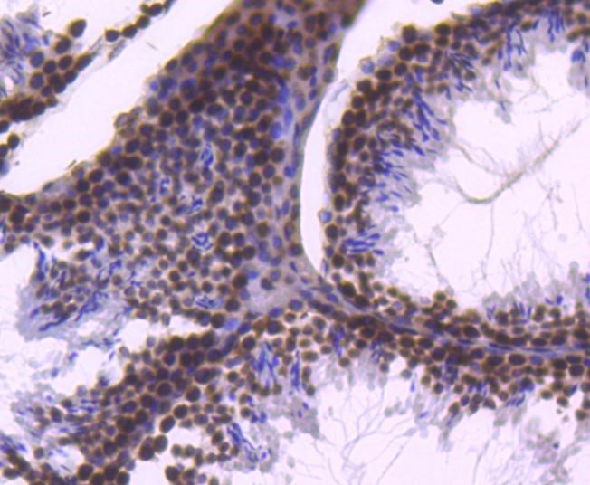
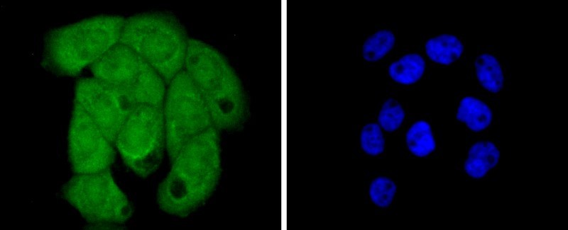
    The nucleosome, made up of four core histone proteins (H2A, H2B, H3, and H4), is the primary building block of chromatin. Originally thought to function as a static scaffold for DNA packaging, histones have now been shown to be dynamic proteins, undergoing multiple types of post-translational modifications, including acetylation, phosphorylation, methylation, and ubiquitination. The p300/CBP histone acetyltransferases acetylate multiple lysine residues in the amino terminal tail of histone H2B (Lys5, 12, 15, and 20) at gene promoters during transcriptional activation. Hyper-acetylation of the histone tails neutralizes the positive charge of these domains and is believed to weaken histone-DNA and nucleosome-nucleosome interactions, thereby destabilizing chromatin structure and increasing the access of DNA to various DNA-binding proteins. In addition, acetylation of specific lysine residues creates docking sites that facilitate recruitment of many transcription and chromatin regulatory proteins that contain a bromodomain, which binds to acetylated lysine residues. Histone H2B is mono-ubiquitinated at Lys120 during transcriptional activation by the RAD6 E2 protein in conjunction with the BRE1A/BRE1B E3 ligase (also known as RNF20/RNF40). Mono-ubiquitinated histone H2B Lys120 is associated with the transcribed region of active genes and stimulates transcriptional elongation by facilitating FACT-dependent chromatin remodeling. In addition, it is essential for subsequent methylation of histone H3 Lys4 and Lys79, two additional histone modifications that regulate transcriptional initiation and elongation. In response to metabolic stress, AMPK is recruited to responsive genes and phosphorylates histone H2B at Lys36, both at promoters and in transcribed regions of genes, and may regulate transcriptional elongation. In response to multiple apoptotic stimuli, histone H2B is phosphorylated at Ser14 by the Mst1 kinase. Upon induction of apoptosis, Mst1 is cleaved and activated by caspase-3, leading to global phosphorylation of histone H2B during chromatin condensation. Interestingly, histone H2B is rapidly phosphorylated at irradiation-induced DNA damage foci in mouse embryonic fibroblasts. In this case, phosphorylation at Ser14 is rapid, depends on prior phosphorylation of H2AX Ser139, and occurs in the absence of apoptosis, suggesting that Ser14 phosphorylation may have distinct roles in DNA-damage repair and apoptosis.
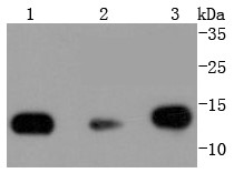
     
    UniProt ID: 
    P33778
    Overview
    Reactivity
    Human, mouse, rat, bovine, chicken, Drosophilia melanogaster, Zebrafish
    Tested applications
    Western Blotting 1:1000
    Immunofluorescence  1:500
    Immunohistochemistry (Paraffin) 1:500
    Immunoprecipitation(IP)  1;100 
    CUT&RUN and CUT&Tag
     
    Optimal dilutions/concentrations should be determined by the end user. 
     
    Specificity
    This antibody recognizes endogenous levels of total histone H2B protein. This antibody does not cross-react with other histone proteins.
    Properties
    Immunogen
    a synthetic peptide corresponding to residues surrounding Val118 of human histone H2B protein.
    Clonality
    Monoclonal, clone number: 4K9
    Isotype
    Rabbit IgG
    Form
    Liquid, 100 μl,1mg/ml, PBS (pH 7.2) and 40% Glycerol,0.02% Sodium Azide
    Storage instruction
    Store at +4°C short term (1-2 weeks). Aliquot and store at -20°C, Avoid freeze / thaw cycle.
    Host
    Rabbit
    Applications
    WB Image

    Western Blot: Histone H2B Antibody - Analysis of Histone H2B on different lysates using anti-Histone H2B antibody at 1/1,000 dilution. 
    Lane 1: Hela Lane 2: NIH/3T3 Lane 3: PC12
     
    IHC Image

    Immunohistochemistry-Paraffin: Histone H2B Antibody - Analysis of paraffin-embedded mouse testis tissue using anti-Histone H2B antibody. Counter stained with hematoxylin
    ICC/IF Image

    Immunocytochemistry/Immunofluorescence: Histone H2B Antibody - Staining Histone H2B in A431 cells (green). The nuclear counter stain is DAPI (blue). Cells were fixed in paraformaldehyde, permeabilised with 0.25% Triton X100/PBS.

    Related Products

    Reviews

    loading...
    Content *:
    Customer Review :
    Verification Code * :
    refresh image

    Shipping and Paying Info
    ......
    Order Information
    You can place an order online step by step as blow.

    Step 1. Register for a new account and Log in, Find out your product by searching our website.
    Step 2. Add the Product into your shopping basket, select your settlement currency.
    Step 3. Checkout by Paypal, Pay by credit card or bank transfer for your order.
    Step 4. Ship the goods for you, providing you package tracking numbers. 

    Any questions, please contact us via email: sale@engibodybio.com
    Payments by
    All our products are For Research Use Only. Not for diagnostic or therapeutic usages.