Histone methyltransferase that specifically trimethylates 'Lys-9' of histone H3. H3 'Lys-9' trimethylation represents a specific tag for epigenetic transcriptional repression by recruiting HP1 (CBX1, CBX3 and/or CBX5) proteins to methylated histones. Mainly functions in euchromatin regions, thereby playing a central role in the silencing of euchromatic genes. H3 'Lys-9' trimethylation is coordinated with DNA methylation. Probably forms a complex with MBD1 and ATF7IP that represses transcription and couples DNA methylation and histone 'Lys-9' trimethylation. Its activity is dependent on MBD1 and is heritably maintained through DNA replication by being recruited by CAF-1. SETDB1 is targeted to histone H3 by TRIM28/TIF1B, a factor recruited by KRAB zinc-finger proteins.
Anti-KMT1E / SETDB1 Rabbit mAb
Catalog number :AT0409
- Overview
- Description
- Rabbit monoclonal antibody to KMT1E / SETDB1
- Reactivity
- Mouse, Human, Rat
- Tested applications
- WB: 1/1000 - 1/10000. Predicted molecular weight: 155 kDa.
IHC-P: Use at an assay dependent dilution.
ICC/IF: Use at an assay dependent concentration. PubMed: 21921037
IP: Use a concentration of 1 - 4 mg/ml.
- Properties
- Immunogen
Immunogen was a synthetic peptide, which represented a portion of human SET domain, bifurcated 1 encoded within exon 19 (LocusLink ID 9869).
- Clonality
- Monoclonal, clone number: 1F7
- Isotype
- Rabbit IgG
- Form
- Liquid
Preservative: 0.1% Sodium Azide
Constituents: 8mM PBS, 60mM Citrate, 150mM Tris, pH 7-8
- Storage instruction
- Shipped at 4°C. Upon delivery aliquot and store at -20°C. Avoid freeze / thaw cycles.
- Database links
- Entrez Gene: 9869 Human
- Entrez Gene: 84505 Mouse
- Omim: 604396 Human
- SwissProt: Q15047 Human
- SwissProt: O88974 Mouse
- Unigene: 643565 Human
- Applications
- IHC Image
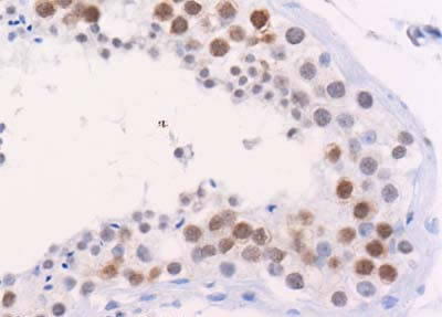
 AT0409 (4ug/ml) staining KMT1E in human testis using an automated system (DAKO Autostainer Plus). Using this protocol there is strong staining of KMT1E in nuclear compartments of the germinal epithelium.
Sections were rehydrated and antigen retrieved with the Dako 3 in 1 AR buffer citrate pH6.1 in a DAKO PT link. Slides were peroxidase blocked in 3% H2O2 in methanol for 10 mins. They were then blocked with Dako Protein block for 10 minutes (containing casein 0.25% in PBS) then incubated with primary antibody for 20 min and detected with Dako envision flex amplification kit for 30 minutes. Colorimetric detection was completed with Diaminobenzidine for 5 minutes. Slides were counterstained with Haematoxylin and coverslipped under DePeX. Please note that, for manual staining, optimization of primary antibody concentration and incubation time is recommended. Signal amplification may be required.
- IP/CoIP Image
 Whole cell lysate (2 mg) from 293T cells that were mock transfected (1 and 2) or transfected with a SETDB1 expression construct (3 and 4). Rabbit anti-SETDB1 antibodies; AT0409 (2 and 4) and a competitor's antiserum (1 and 3) were used at 1 mcg/mg lysate for IP. AT0409 was used at 0.2 mg/ml for Western Blot Detection: Chemiluminescence with exposure times of 1 to 5 minutes.