MGMT (O6 methylguanine DNA methyltransferase) is an enzyme that transfers methyl groups from O(6) methylguanine, and other methylated moities of DNA, to a cysteine residue in itself, thus repairing DNA damage caused by alkylation in a single step reaction. This is a suicide reaction as the enzyme is irreversibly inactivated. MGMT prevents mutagenesis and malignant transformation, and also provides resistance to chemotherapy with alkylating agents among cancer patients.
Anti-MGMT Rabbit mAb
Catalog number :AT0408
- Overview
- Description
- Rabbit monoclonal antibody to MGMT
- Reactivity
- Human, Mouse, Rat
- Tested applications
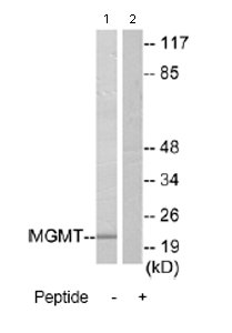
- WB: 1/500 - 1/1000. Detects a band of approximately 22 kDa (predicted molecular weight: 22 kDa).
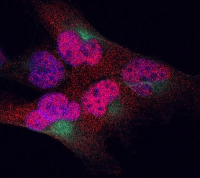
ICC/IF: Use at an assay dependent concentration.
ELISA: 1/5000.
- Properties
- Immunogen
Synthetic peptide derived from the internal region of human MGMT.
- Clonality
- Monoclonal, clone number: 4B23
- Isotype
- Rabbit IgG
- Form
- Liquid
Preservative: 0.02% Sodium Azide
Constituents: 50% Glycerol, PBS, 150mM Sodium chloride, pH 7.4
- Storage instruction
- Shipped at 4°C. Upon delivery aliquot and store at -20°C. Avoid freeze / thaw cycles.
- Database links
- Entrez Gene: 4255 Human
- Entrez Gene: 25332 Rat
- Omim: 156569 Human
- SwissProt: P16455 Human
- SwissProt: P24528 Rat
- Unigene: 501522 Human
- Unigene: 9836 Rat
- Applications
- WB Image
 All lanes : Anti-MGMT antibody (AT0408) at 1/500 dilution
Lane 1 : Extracts from Jurkat cells.
Lane 2 : Extracts from Jurkat cells with immunising peptide at 5 µg
Lysates/proteins at 5 µg per lane.
Predicted band size : 22 kDa
Observed band size : 22 kDa
- IHC Image
 AT0408 staining MGMT in T98G human glioma cell line by Immunocytochemistry/ Immunofluorescence. Cells were fixed in paraformaldehyde and permeabilized in 0.1% Triton X-100 prior to blocking in 3% BSA for 30 minutes at 20°C. The primary antibody was diluted 1/100 and incubated with the sample for 1 hour at 20°C. The secondary antibody was Alexa Fluor® 555-conjugated goat anti-rabbit polyclonal, diluted 1/300. MGMT staining is shown in red (AF 555) and stains nuclei and to a small extent, cytoplasm. Nuclei were stained with DAPI (blue), while microtubules were stained using an anti-tubulin FITC conjugate (green).