NF-kappa-B is a pleiotropic transcription factor which is present in almost all cell types and is involved in many biological processed such as
inflammation, immunity, differentiation, cell growth, tumorigenesis and apoptosis. NF-kappa-B is a homo- or heterodimeric complex formed by
the Rel-like domain-containing proteins RELA/p65, RELB, NFKB1/p105, NFKB1/p50, REL and NFKB2/p52 and the heterodimeric p65-p50 complex appears to be most abundant one. The dimers bind at kappa-B sites in the DNA of their target genes and the individual dimers have distinct preferences for different kappa-B sites that they can bind with distinguishable affinity and specificity. Different dimer combinations act as transcriptional activators or repressors, respectively. NF-kappa-B is controlled by various mechanisms of post-translational modification and subcellular compartmentalization as well as by interactions with other cofactors or corepressors. NF-kappa-B complexes are held in the cytoplasm in an inactive state complexed with members of the NF-kappa-B inhibitor (I-kappa-B) family. In a conventional activation pathway, I-kappa-B is phosphorylated by I-kappa-B kinases (IKKs) in response to different activators, subsequently degraded thus liberating the active NF-kappa-B complex which translocates to the nucleus. NF-kappa-B heterodimeric p65-p50 and RelB-p50 complexes are transcriptional activators. The NF-kappa-B p50-p50 homodimer is a transcriptional repressor, but can act as a transcriptional activator when associated with BCL3. NFKB1 appears to have dual functions such as cytoplasmic retention of attached NF-kappa-B proteins by p105 and generation of p50 by a cotranslational processing. The proteasome-mediated process ensures the production of both p50 and p105 and preserves their independent function, although processing of NFKB1/p105 also appears to occur post-translationally. p50 binds to the kappa-B consensus sequence 5'-GGRNNYYCC-3', located in the enhancer region of genes involved in immune response and acute phase reactions. In a complex with MAP3K8, NFKB1/p105 represses MAP3K8-induced MAPK signaling; active MAP3K8 is released by proteasome-dependent degradation of NFKB1/p105.
Anti-NF-kB p105 / p50 Rabbit mAb - ChIP, CUT&RUN and CUT&Tag Grade
Catalog number :AT0362
- Overview
- Description
- Rabbit monoclonal antibody to NFkB p105 / p50
- Reactivity
- Mouse, Rat, Human
- Tested applications
ChIP, CUT&RUN and CUT&TagIHC-Fr : Use at an assay dependent dilution. IP : Use at an assay dependent dilution. WB : 1/400. Predicted molecular weight: 50 kDa. ChIP : Use at an assay dependent dilution. Flow Cyt : Use at an assay dependent concentration. EMSA : Use at an assay dependent concentration. IHC-P : 1/100. ICC/IF : Use at an assay dependent concentration.
- Properties
- Immunogen
Synthetic peptide derived from the region aa330 to aa433 of the Human NFkB p50 subunit.
- Clonality
- Monoclonal, clone number: 6HS7
- Isotype
- IgG
- Form
- Liquid
Preservative: 0.1% Sodium azide
Constituents: PBS, 0.2% Gelatin
- Storage instruction
- Store at +4°C short term (1-2 weeks). Store at -20°C or -80°C. Avoid freeze / thaw cycle.
- Applications
- WB Image
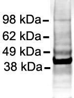 Application Western blot
SampleHuman Cell lysate - whole cell (iPS derived neuronal cells)Loading amount 20 µgSpecificationiPS derived neuronal cellsGel Running ConditionsNon-reduced Denaturing (4-12%)Blocking stepMilk as blocking agent for 1 hour(s) and 0 minute(s) · Concentration: 5% · Temperature: 25°C
Application Western blot
SampleHuman Cell lysate - whole cell (iPS derived neuronal cells)Loading amount 20 µgSpecificationiPS derived neuronal cellsGel Running ConditionsNon-reduced Denaturing (4-12%)Blocking stepMilk as blocking agent for 1 hour(s) and 0 minute(s) · Concentration: 5% · Temperature: 25°COther product details
Application Western blotDilution1/400Incubation time16 hour(s) and 0 minute(s) · Temperature: 4°C · Diluent: 3% BSA in TBS-TSecondary antibody
Application Western blotIRDye 680 conjugated IgG (Licor Biosciences)
Dilution1/5000Detection
Application Western blotDetection methodOdisseyExposure1 second(s)BandsSpecific: 50 kDa Non-specific: many kDa
- ICC/IF Image
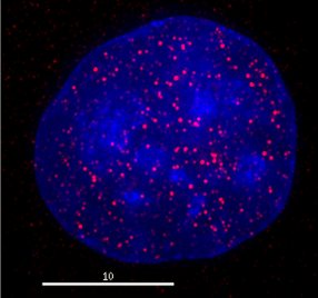 HeLa cells were fixed with 4% formaldehyde in PEM buffer. The coverslip was incubated in blocking buffer of 5% powdered milk in TBS-T plus 0.02% sodium azide for 1 hour at room temperature. Blocking buffer was removed and primary antibody was added at a dilution of 1/500 and incubated overnight at 4 degrees celsius. The coverslips were then washed 4-5 times with blocking buffer for 5 minutes. Secondary antibody, goat anti-rabbit Alexa 594, was added at a dilution of 1/1000 and incubated at room temperature for one hour. From this point on coverslips were covered with foil to protect them from light. They were washed 5 times with TBS-T and then one time with PEM, for 5 minutes each wash. The coverslips were fixed 10-30 minutes in 4% formaldehyde in PEM buffer, then washed 3 times with PEM buffer for 5 minutes. 0.1M ammonium chloride in PEM buffer was added for 10 minutes to quench auto-florescence, and then slips were washed 2 times for 5 minutes in PEM followed by 3 washes for 5 minutes in TBS-T. Coverslips were then counterstained with DAPI in TBS-T for 1-2 minutes, TBS-T was then added and the coverslips mounted. Red indicates staining by ab7971, blue staining by DAPI.