Anti-PARP1 Rabbit mAb
Catalog number :AT0396
PARP, a 114 kDa nuclear poly (ADP-ribose) polymerase, appears to be involved in DNA repair in response to environmental stress. This protein can be cleaved by many ICE-like caspases in vitro and is one of the main cleavage targets of caspase-3 in vivo. In human PARP, the cleavage occurs between Asp214 and Gly215, which separates the PARP amino-terminal DNA binding domain (24 kDa) from the carboxy-terminal catalytic domain (89 kDa). PARP helps cells to maintain their viability; cleavage of PARP facilitates cellular disassembly and serves as a marker of cells undergoing apoptosis
- Overview
- Description
- Rabbit Monoclonal antibody to PARP1
- Reactivity
- Human; Mouse; monkey; dog; rabbit; Chinese hamster
- Tested applications
- WB: 1/1000 - 1/2000; IF: 1/100-1/500
- Properties
- Immunogen
This antibody is produced by immunizing rabbit with a polypeptides corresponding to surface epitope of human PARP1
- Clonality
- Monoclonal, clone number: 5G1
- Isotype
Rabbit Polyclonal
- Form
- Liquid
Preservative: 0.1% Sodium Azide
Constituents: 0.2% Gelatin, PBS
- Storage instruction
- Store at +4°C short term (1-2 weeks). Store at -20°C or -80°C. Avoid freeze / thaw cycle.
- Database links
- Entrez Gene: 142 Human
- Entrez Gene: 11545 Mouse
- Entrez Gene: 25591 Rat
- Omim: 173870 Human
- SwissProt: P09874 Human
- SwissProt: P11103 Mouse
- SwissProt: P27008 Rat
- Unigene: 177766 Human
- Applications
- WB Image

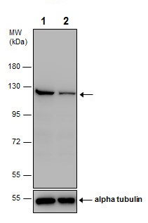
All lanes: Anti-PARP1 antibody at 1/2000 dilutionLane 1: Non-transfected HEK-293T (human epithelial cell line from embryonic kidney transformed with large T antigen) whole cell extractLane 2: PARP1 shRNA transfected HEK-293T (human epithelial cell line from embryonic kidney transformed with large T antigen) whole cell extractLysates/proteins at 30 µg per lane.SecondaryAll lanes: HRP-conjugated anti-rabbit IgGPredicted band size: 113 kDa
- IHC Image

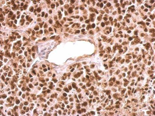
Paraffin-embedded HeLa xenograft tissue stained for PARP1 using this antibody at 1/500 dilution in immunohistochemical analysis.
- IP/CoIP Image

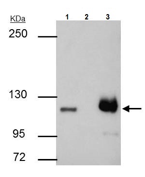
PARP1 was immunoprecipitated from HCT 116 (human colorectal carcinoma cell line) whole cell extract with 4 µg this antibody. Western blot was performed from the immunoprecipitation using this antibody at 1/500 dilution. Anti-Rabbit IgG was used as a secondary reagent.Lane 1: HCT 116 whole cell extract 30 μg.Lane 2: Control IP in HCT 116 whole cell extract with 4 μg of Rabbit IgG.Lane 3: This antibody IP in HCT 116 whole cell extract.
Related Products
Reviews
loading...