Anti-SOX2 Rabbit mAb - ChIP, CUT&RUN and CUT&Tag Grade
Catalog number :AT0700
SOX2 (SRY-RELATED HMG-BOX GENE 2), the basic helix-loop-helix transcription factor, is indispensable for maintaining embryonic stem (ES) cell pluripotency. The gene expresses in self-renewing progenitor cells and acts to inhibit neuronal differentiation. SOX2, along with other transcription factors Oct3/4, klf4, and c-Myc are used to generate induced pluripotent stem cells from mouse fibroblasts by retrovirus mediated introduction. It is expressed throughout the human brain, including the developing hypothalamus, as well as the Rathke pouch, the developing anterior pituitary and the eye. It is necessary for the normal development of the hypothalamo-pituitary and reproductive axes in mice. Mutation of the SOX2 gene in progenitor cells leads to cerebral malformation, severe eye defects, and hypothalamo-pituitary abnormalities.
- Overview
- Reactivity
- Mouse, Rat, Human, Sheep
- Tested applications
- WB (1:500-1:1000), IHC (1:50-1:200), Immunofluorescence (1:50-1:250), Flow Cytometry (1:100), ChIP, CUT&RUN and CUT&Tag
- Specificity
- This antibody detects endogenous levels of SOX2.
- Properties
- Immunogen
- A synthetic peptide made to the N-terminal region of human SOX2 protein (within residues 1-100). [Swiss-Prot# P48431]
- Clonality
- Monoclonal, clone number: 52B9
- Isotype
- Rabbit IgG
- Storage instruction
- Store at +4°C short term (1-2 weeks). Aliquot and store at -20°C
- Applications
- WB Image
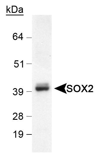
Western Blot: SOX2 Antibody- Detection of SOX2 in mouse brain lysate using AT0700 (1:1000).
- IHC Image
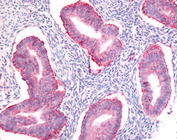
Immunohistochemistry-Paraffin: SOX2 Antibody - Staining of human uterus, endometrial glands.
- ICC/IF Image
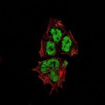
Immunofluorescence: SOX2 - Analysis of NTERA-2 cells using SOX2 pAb (green).Red: Actin filaments have been labeled with Alexa Fluor-555 phalloidin.
- Application Image
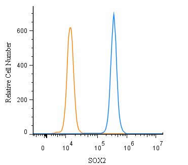
Flow Cytometry: SOX2 Antibody- An intracellular stain was performed on A549 with SOX2 Antibody and a matched isotype control.Cells were fixed with 4% PFA and then permeablized with 0.1% saponin. Cells were incubated in an antibody dilution of 1:500 for 30 minutes at room temperature, followed by Rabbit IgG (H+L) Cross-Adsorbed Secondary Antibody, Dylgiht 550 (SA5-10033, Thermo Fisher).
Related Products
Reviews
loading...