Anti-SQSTM1 / p62 Rabbit mAb
Catalog number :AT1793
Sequestosome 1 (SQSTM1, p62) is a ubiquitin binding protein involved in cell signaling, oxidative stress, and autophagy. It was first identified as a protein that binds to the SH2 domain of p56Lck and independently found to interact with PKCζ. SQSTM1 was subsequently found to interact with ubiquitin, providing a scaffold for several signaling proteins and triggering degradation of proteins through the proteasome or lysosome. Interaction between SQSTM1 and TRAF6 leads to the K63-linked polyubiquitination of TRAF6 and subsequent activation of the NF-κB pathway. Protein aggregates formed by SQSTM1 can be degraded by the autophagosome. SQSTM1 binds autophagosomal membrane protein LC3/Atg8, bringing SQSTM1-containing protein aggregates to the autophagosome. Lysosomal degradation of autophagosomes leads to a decrease in SQSTM1 levels during autophagy; conversely, autophagy inhibitors stabilize SQSTM1 levels. Studies have demonstrated a link between SQSTM1 and oxidative stress. SQSTM1 interacts with KEAP1, which is a cytoplasmic inhibitor of NRF2, a key transcription factor involved in cellular responses to oxidative stress. Thus, accumulation of SQSTM1 can lead to an increase in NRF2 activity.
- Overview
- Reactivity
- Mouse, Rat, Human
- Tested applications
- WB : 1/500-1/2000.ICC/IF : 1/50-1/200.Not yet tested in other applications. Optimal dilutions/concentrations should be determined by the end user.
- Specificity
- Recognizes endogenous levels of total SQSTM1/p62.
- Properties
- Immunogen
- A synthetic peptide made to an internal region of the human p62/SQSTM1 protein (within residues 350-400).
- Clonality
- Monoclonal, clone number: 4D11
- Isotype
- Rabbit IgG
- Storage instruction
- Store at +4°C short term (1-2 weeks). Aliquot and store at -20°C
- Database links
- Q13501
- Applications
- WB Image

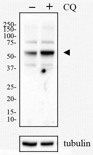
Western Blot: p62/SQSTM1 Antibody - HeLa cells were treated with (+) or without 50 uM (-) of Chloriquine (CQ) for 24 hours. Total cell lysates were prepared and separated on a 12% gel by SDS-PAGE. Protein was transferred to PVDF membrane and blocked in 5% non-fat milk. The membrane was then probed with anti-p62/SQSMT1 antibody (1:1000) in 5% milk and detected with an anti-rabbit HRP secondary antibody using chemiluminescence. Note the upregulation of p62 (arrowhead) in response to chloroquine treatment and the blockage of autophagy.
- ICC/IF Image

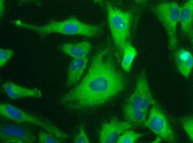
Immunocytochemistry/Immunofluorescence: p62/SQSTM1 Antibody - SQSTM1 staining in of HeLa cells detected with a Dylight 488 labeled secondary antibody (Green) with Hoechst 33258 nuclear counterstain (Blue).
Related Products
Reviews
loading...