Anti-Smad4 Rabbit mAb - ChIP, CUT&RUN and CUT&Tag Grade
Catalog number :AT0647
Members of the Smad family of signal transduction molecules are components of a critical intracellular pathway that transmits TGF-β signals from the cell surface into the nucleus. Three distinct classes of Smads have been defined: the recepter-regulated Smads (R-Smads), which include Smad1, 2, 3, 5, 8; the common-mediator Smad (co-Smad), Smad4; and the antagonistic or inhibitory Smads (I-Smads), Smad6 and 7. Briefly, activated type I receptors associate with specific R-Smads and phosphorylate them on a conserved SSXS motif at the carboxy-terminus of the proteins. The phosphorylated R-Smad dissociates from the receptor and forms a heteromeric complex with the co-Smad, Smad4, and together the complex moves to the nucleus. Once in the nucleus, Smads can target a variety of DNA binding proteins to regulate transcriptional responses.
- Overview
- Reactivity
- Mouse, Rat, Human
- Tested applications
- Western Blotting 1:2000Immunoprecipitation 1:100Immunohistochemistry (Paraffin) 1:500Immunofluorescence (Immunocytochemistry) 1:300Flow Cytometry 1:200ChIP, CUT&RUN and CUT&Tag
- Specificity
- This antibody detects endogenous levels of total Smad4 protein.
- Properties
- Immunogen
- Synthetic peptide conjugated to KLH derived from within residues 150 - 250 of Human Smad4.
- Clonality
- Monoclonal, clone number: 3DA7
- Isotype
- Rabbit IgG
- Form
- Liquid, 100 μl,1mg/ml, PBS (pH 7.2) and 40% Glycerol,0.02% Sodium Azide
- Storage instruction
- Store at +4°C short term (1-2 weeks). Aliquot and store at -20°C, Avoid freeze / thaw cycle.
- Host
- Rabbit
- Applications
- WB Image
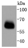
Western Blot:Smad4 Antibody - Analysis of Smad4 on PC-12 cells lysates using anti-Smad4 antibody at 1/1,000 dilution.
- IHC Image
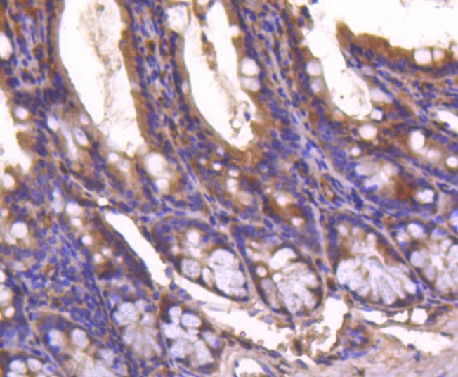
Immunohistochemistry-Paraffin:Smad4 Antibody - Analysis of paraffin-embedded mouse colon tissue using anti-Smad4 antibody. Counter stained with hematoxylin.
- ICC/IF Image
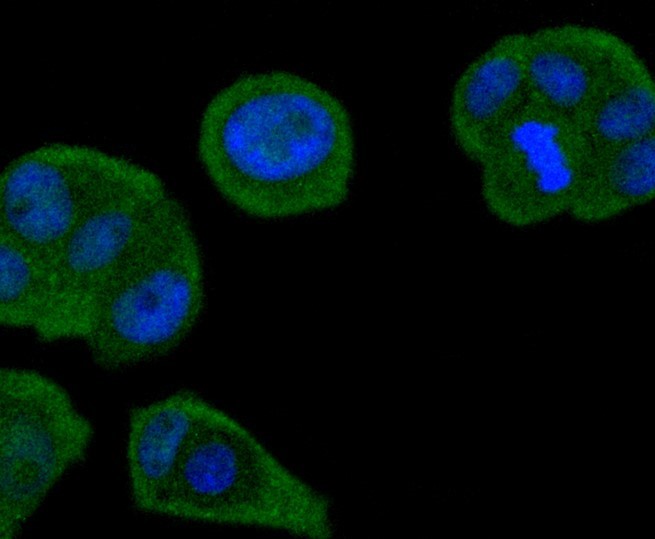
Immunocytochemistry/Immunofluorescence:Smad4 Antibody - Staining Smad4 in MCF-7 cells (green). The nuclear counter stain is DAPI (blue). Cells were fixed in paraformaldehyde, permeabilised with 0.25% Triton X100/PBS.
Related Products
Reviews
loading...