Anti-c-Fos Rabbit mAb - ChIP, CUT&RUN and CUT&Tag Grade
Catalog number :AT0631
The Fos family of nuclear oncogenes includes c-Fos, FosB, Fos-related antigen 1 (FRA1), and Fos-related antigen 2 (FRA2). While most Fos proteins exist as a single isoform, the FosB protein exists as two isoforms: full-length FosB and a shorter form, FosB2 (Delta FosB), which lacks the carboxy-terminal 101 amino acids. The expression of Fos proteins is rapidly and transiently induced by a variety of extracellular stimuli including growth factors, cytokines, neurotransmitters, polypeptide hormones, and stress. Fos proteins dimerize with Jun proteins (c-Jun, JunB, and JunD) to form Activator Protein-1 (AP-1), a transcription factor that binds to TRE/AP-1 elements and activates transcription. Fos and Jun proteins contain the leucine-zipper motif that mediates dimerization and an adjacent basic domain that binds to DNA. The various Fos/Jun heterodimers differ in their ability to transactivate AP-1 dependent genes. In addition to increased expression, phosphorylation of Fos proteins by Erk kinases in response to extracellular stimuli may further increase transcriptional activity. Phosphorylation of c-Fos at Ser32 and Thr232 by Erk5 increases protein stability and nuclear localization. Phosphorylation of FRA1 at Ser252 and Ser265 by Erk1/2 increases protein stability and leads to overexpression of FRA1 in cancer cells. Following growth factor stimulation, expression of FosB and c-Fos in quiescent fibroblasts is immediate, but very short-lived, with protein levels dissipating after several hours. FRA1 and FRA2 expression persists longer, and appreciable levels can be detected in asynchronously growing cells. Deregulated expression of c-Fos, FosB, or FRA2 can result in neoplastic cellular transformation; however, Delta FosB lacks the ability to transform cells
- Overview
- Reactivity
- Mouse, Rat, Human
- Tested applications
- Western Blotting 1/500~1/2000Immunofluorescence 1/100~1/500Flow Cytometry 1/20~1/100IP 1/50ChIP, CUT&RUN and CUT&Tag
- Specificity
- This Antibody detects endogenous levels of total c-Fos protein. The antibody does not cross-react with other Fos proteins, including FosB, FRA1 and FRA2.
- Properties
- Immunogen
- Recombinant human c-Fos protein
- Clonality
- Monoclonal, clone number: 5DB7
- Isotype
- Rabbit IgG
- Form
- Liquid, 100 μl,1mg/ml
- Storage instruction
- store at -20°C
- Host
- Rabbit
- Applications
- WB Figure 1
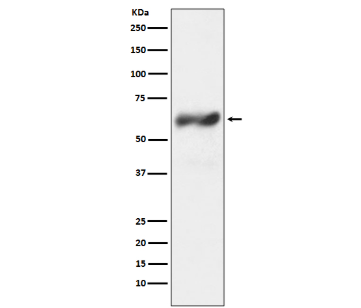
Western blot analysis of c-Fos expression in HeLa cell lysate treated
with TPA. Predicted molecular weight: 41kDa.
Observed molecular weight: 62 kDa
Recommended secondary antibody: Goat anti rabbit IgG-HRP(Engibody, Catalog number:AT0097)
Recommended ECL: ECL Pico-Detect™ Western ChemiluminescentHRP Substrate (Engibody, Catalog number: IF6747)
- WB Figure 2
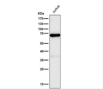
Western Blot: c-Fos Antibody - Analysis on Jurkat lysate prepared inRIPA buffer containing 1% Triton-X100; 1% SDS and 1% SDC. Blocking: 1% LFDM for 30 min at RT; primary antibody dilution 1:1000 incubated at 4°C overnight.
Predicted molecular weight: 41kDa.
Observed molecular weight: 62 kDa
Recommended secondary antibody: Goat anti rabbit IgG-HRP(Engibody, Catalog number:AT0097)
Recommended ECL: ECL Pico-Detect™ WesternChemiluminescent HRP Substrate (Engibody, Catalog number:IF6747)
- ICC/IF Image
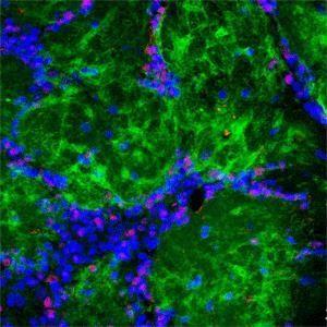
Immunofluorescence: c-Fos Antibody - Rabbit antibody to cFos (c-fos) at a 1: 500 dilution and DAPI counterstained appearing in blue.
Related Products
Reviews
loading...