Anti-c-Jun Rabbit mAb - ChIP, CUT&RUN and CUT&Tag Grade
Catalog number :AT0632
c-Jun is a member of the Jun family containing c-Jun, JunB, and JunD, and is a component of the transcription factor activator protein-1 (AP-1). AP-1 is composed of dimers of Fos, Jun, and ATF family members and binds to and activates transcription at TRE/AP-1 elements. Extracellular signals including growth factors, chemokines, and stress activate AP-1-dependent transcription. The transcriptional activity of c-Jun is regulated by phosphorylation at Ser63 and Ser73 through SAPK/JNK. Knock-out studies in mice have shown that c-Jun is essential for embryogenesis, and subsequent studies have demonstrated roles for c-Jun in various tissues and developmental processes including axon regeneration, liver regeneration, and T cell development. AP-1 regulated genes exert diverse biological functions including cell proliferation, differentiation, and apoptosis, as well as transformation, invasion and metastasis, depending on cell type and context. Other target genes regulate survival, as well as hypoxia and angiogenesis. Research studies have implicated c-Jun as a promising therapeutic target for cancer, vascular remodeling, acute inflammation, and rheumatoid arthritis.
- Overview
- Reactivity
- Mouse, Rat, Human, Monkey
- Tested applications
- Western Blotting 1:1000Immunoprecipitation 1:50Immunohistochemistry (Paraffin) 1:300Immunofluorescence (Immunocytochemistry) 1:400Flow Cytometry 1:100ChIP, CUT&RUN and CUT&Tag
- Specificity
- This Antibody detects endogenous levels of total c-Jun protein, regardless of phosphorylation state.
- Properties
- Immunogen
- Purified recombinant fragment of human c-Jun expressed in E. Coli.
- Clonality
- Monoclonal, clone number: 5G1
- Isotype
- Rabbit IgG
- Form
- Liquid, 100 μl,1mg/ml
- Storage instruction
- Store at +4°C short term (1-2 weeks). Aliquot and store at -20°C
- Host
- Rabbit
- Applications
- WB Image
-

Western Blot: c-jun Antibody - Western blot analysis using c-Jun Antibody against NIH/3T3 cell lysate.
Western Blot: c-jun Antibody - Western blot analysis using c-Jun Antibody against NIH/3T3 cell lysate.
- IHC Image

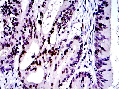
Immunohistochemistry: c-jun Antibody - Immunohistochemical analysis of paraffin-embedded colon cancer tissues using c-Jun antibody with DAB staining.
- ICC/IF Image

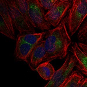
Immunocytochemistry/Immunofluorescence: c-jun Antibody - Immunofluorescence analysis of Hela cells using c-Jun antibody (green). Blue: DRAQ5 fluorescent DNA dye. Red: Actin filaments have been labeled with Alexa Fluor-555 phalloidin.
Related Products
Reviews
loading...