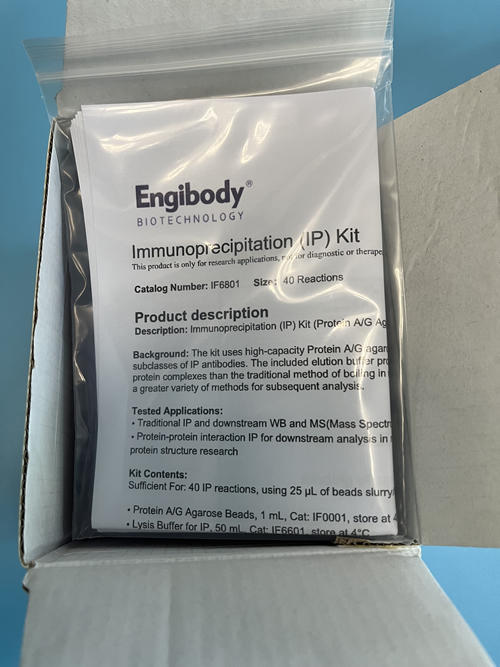
The kit uses high-capacity Protein A/G agarose affinity Beads for efficient binding of most species and subclasses of IP antibodies. The included IgG elution buffer provides milder and less denaturing recovery of antibody:antigen complexes than the traditional method of boiling in reducing sample loading buffer for SDS-PAGE, facilitating a greater variety of methods for subsequent analysis.
IP Kit
Catalog number :IF6801
- Overview
- Description
- Immunoprecipitation(IP) Kit (Protein A/G Agarose Beads)
- Tested applications
- • Traditional IP and downstream WB• IP for downstream analysis in nonreducing conditions, such as enzyme activity assay, protein structure research
- Product Picture

- Product Picture 1

- Product Picture 2
- Product Picture 3
- Properties
- Components
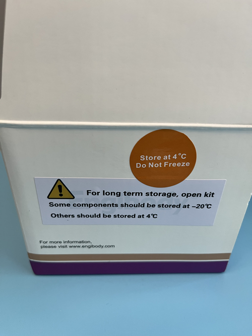
- Sufficient For: 40 IP reactions, using 25 µL of beads slurry/reaction• Protein A/G Agarose Beads, 1 mL, Cat: IF0001, store at 4°C• Lysis Buffer for IP, 50 mL, Cat: IF6601, store at 4°C• Wash Buffer for IP, 50 mL, Cat: IF9210, store at 4°C• Elution Buffer, 4 mL, Cat: IF9213, store at 4°C• Neutralization Buffer, 0.5 mL, Cat: IF9216, store at 4°C• Sample Loading Buffer (Reducing and Denaturing, 5X), 1 mL, Cat: IF6740, store at -20°C• Normal Rabbit IgG (Whole Molecule) Isotype Control,100 μL, Cat: AT1597, store at -20°C• Normal Mouse IgG (Whole Molecule) Isotype Control,100 μL, Cat: AT1596, store at -20°C
- Applications
- Highlights
- • Fast—immunoprecipitate (IP) in less than one hour• Mild elution conditions—recover antigen without harsh detergents or reducing agents• Easy-to-follow instructions—purify target protein from crude lysate in four simple steps• Suitable for most common antibody types—Protein A/G provides excellent binding for most mouse, rabbit, human and goat IgG subclasses• Complete kit—includes lysis buffer, wash buffer, and elution buffers, sample loading buffer
- Work Flow Image

- Protocols
- Procedure for IPA. Mammalian Cell LysisProtocol I: Lysis of Adherent Cell Cultures1. Carefully remove culture medium from cells.2. Wash the cells once with 1X PBS.3. Add ice-cold Lysis Buffer for IP (Cat: IF6601) to the cells. Incubate on ice for 10-20 minutes with periodic mixing.Note: Protease/Phosphatase Inhibitor Cocktail should be added to Lysis Buffer. Protease inhibitors are rapidly inactivated in aqueous solutions and should be added to solutions immediately before use.Table 1. Suggested volume of IP Lysis Buffer to use for different standard culture plates.Plate Size/Surface AreaVolume of Lysis Buffer for IP100 mm dish 500-1000 μL60 mm dish 250-500 μL4. Transfer the lysate to a microcentrifuge tube and centrifuge at ~ 13,000 × g for 10 minutes to pellet the cell debris.5. Transfer supernatant to a new tube for protein concentration determination and further analysis.Protocol II: Lysis of Cell Suspension Cultures1. Centrifuge the cell suspension at 1000 × g for 5 minutes to pellet the cells. Discard the supernatant.2. Wash cells once by suspending the cell pellet in PBS. Centrifuge at 1000 × g for 5 minutes to pellet cells.3. Add ice-cold Lysis Buffer for IP (Cat: IF6601) to the cell pellet. Use 500 μL of Lysis Buffer for IP per 50 mg of wet cell pellet (i.e., 10:1 v/w). If using a large amount of cells, first add 10% of the final volume of Lysis Buffer for IP to the cell pellet and pipette the mixture up and down to mix. Add the remaining volume of Lysis Buffer for IP to the cell suspension.Note: Protease/Phosphatase Inhibitor Cocktail should be added to Lysis Buffer. Protease inhibitors are rapidly inactivated in aqueous solutions and should be added to solutions immediately before use.4. Incubate lysate on ice for 5 minutes with periodic mixing. Remove cell debris by centrifugation at ~ 13,000 × g for 10 minutes. Transfer supernatant to a new tube for protein concentration determination and further analysis.B. Pre-clear lysate using the Protein A/G agarose beads (this step is optional, pre-clearing the lysate will reduce non-specific binding of proteins to the agarose beads. You can do this step if high background occurs in downstream assay.)1. Pre-clear the cell lysate by adding 20 µL of Protein A/G agarose beads slurry per 1 mL of cell lysate and incubating at 4 °C for 10 minutes on a rotator.2. Remove the Protein A/G agarose beads by centrifugation at 14,000xg at 4°C for 5 minutes. Transfer the supernatant (cell lysate) to a fresh centrifuge tube on ice.Note: In this step after centrifugation the Protein A/G agarose beads should be discarded, the supernatant should be saved for further IP.3. Determine the protein concentration of the cell lysate (e.g. if performing a Bradford assay one must dilute the cell lysate at least 1:10 before determining the protein concentration because of the interference of the detergents in the lysis buffer with the Coomassie-based reagent).C. IPNote:• Protein A/G Agarose beads (Cat: IF0001) should be resuspended well before used.• Perform all IP steps at 4°C unless otherwise indicated.• The amount of antibody-antigen complex required and incubation time depends upon the system used and the affinity of the antibody, protein interactions and must be optimized for each system.• It may be necessary to optimize the binding time for each application1. Transfer 1 mL of the above cell lysate, or approximately 1000 µg total cellular protein, to a 1.5 mL microcentrifuge tube. Add the recommended volume of the primary antibody (optimal antibody concentration should be determined by antibody datasheet) and Normal IgG(2.5µL), then incubate for 2 hours or overnight at 4 °C on a rotator.Note: Normal Rabbit IgG (Cat: AT1597) is a nonspecific antibody, it can be used as Isotype Control of primary antibody from rabbit. Normal Mouse IgG (Cat: AT1596) is also a nonspecific antibody, it can be used as Isotype Control of primary antibody from mouse.2. Add 25 µL-40 µL of resuspended volume of protein A/G agarose beads slurry to capture the antigen-antibody immunocomplex. Incubate at 4 °C on a rocker platform or rotating device for 1-2 hours.3. Collect immunoprecipitation complex by centrifugation at 2,500 rpm (approximately 1,000xg) for 5 minutes at 4°C. Carefully aspirate and discard supernatant.4. Wash the complex 2-3 times with 0.5 mL Wash buffer for IP (Cat: IF9210), each washing lasts for 5-10 minutes on a gentle rotating device and repeat centrifugation step above.Tip: Increase the number of washes to 5-6 times if high background occurs.D. Elution and obtaining of the antigen proteinTwo methods are recommended according to protein characteristics or further usage:Option A: Elution with Elution Buffer (Cat: IF9213). This is a fast and efficient elution method. Equilibration of the eluted proteins with Neutralizing Buffer (Cat: IF9216) may help preserve its activity.Option B: Boil with Sample Loading Buffer (Cat: IF6740), Centrifuge and get the supernatants for gel electrophoresis and western blotting.Option A: Elution with Elution Buffer (Non-denaturing elution)The procedure should be performed at room temperature.Note: Do not leave the beads in this buffer more than 20 minutes.1. Add 100 µL of Elution Buffer to each sample and control beads complex.2. Incubate the samples and controls with gentle shaking for 5 minutes at room temperature.3. Centrifuge the beads complex for 60 seconds at 8,000×g.4. Transfer the supernatants to fresh test tubes. Be careful not to transfer any beads.5. In order to equilibrate the eluant, immediately add 10 µL of Neutralizing Buffer to each sample. Then use a pH meter to make sure the pH is 7-8.6. For immediate use, store the eluted proteins at 2–8 °C. Store at –20 °C for long term storage.Option B: Boil with Sample Loading Buffer working solution, Centrifuge and get the supernatants for gel electrophoresis and western blotting. (Denaturing elution)The procedure should be performed at room temperature. Sample Loading Buffer should be at room temperature before use.1. Prepare Sample Loading Buffer working solution, adding 6 µL 5×Sample Loading Buffer into 24 µL Lysis Buffer for IP.2. Add 30 µL of Sample Loading Buffer working solution to each sample and control beads complex.3. Boil the samples and controls for 10 minutes to dissociate the immunocomplexes from the beads.4. Centrifuge the samples and controls at 8,000×g for 60 seconds to pellet any undissolved agarose beads. Transfer the supernatants to fresh test tubes. The samples and controls are ready for loading on SDS-PAGE and immunoblotting.
Related Products
| Catalog number | IF9055 |
| Catalog number | IF6802 |
| Catalog number | IF9058 |
Reviews
loading...